Antibodies
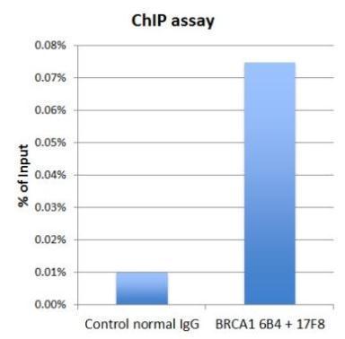
- 5 imagesBRCA1 antibody [17F8] - ChIP grade [GRP85]
ChIP, ELISA, ICC, IF, IHC-P, IP, WB
Human, Mouse
Mouse
Monoclonal
100 μl -
- 7 imagesBRCA1 antibody [6B4] - ChIP grade [GRP86]
ChIP, ICC, IF, IHC-P, IP, WB
Human, Mouse
Mouse
Monoclonal
100 μl -
- 5 imagesMre11 antibody [12D7] [GRP88]
ELISA, FA, ICC, IF, IHC-P, IP, WB
Human, Mouse, Rat
Mouse
Monoclonal
100 μl -
- 12 imagesRad50 antibody [13B3] [GRP89]
ICC, IF, IHC-P, IP, WB
Human, Mouse, Rat, Monkey
Mouse
Monoclonal
100 μl -
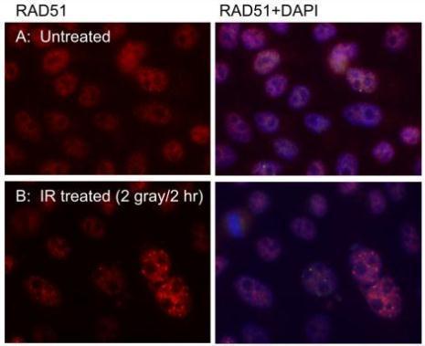
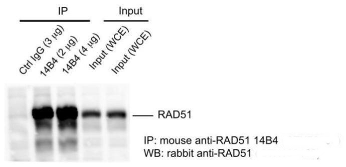
- 15 imagesRad51 antibody [14B4] [GRP90]
ICC, IF, IHC-P, IP, WB
Human, Mouse, Rat, Chicken
Mouse
Monoclonal
100 μl -
- 8 imagesApolipoprotein E antibody [C2C3], C-term [GRP94]
ELISA, ICC, IF, IHC-P, IP, WB
Human
Rabbit
Polyclonal
100 μl -
- 7 images
-
- 6 images
-
- 9 images
-
- 21 imagesSOX2 antibody [N1C3] [GRP105]
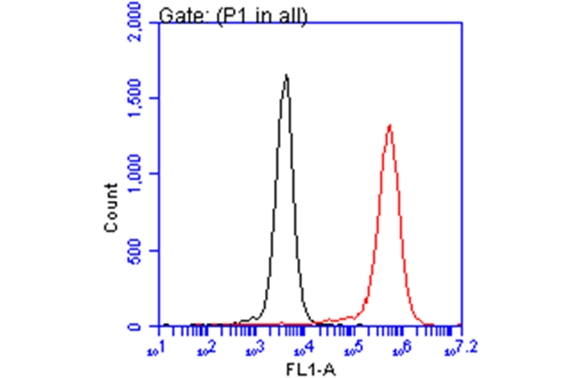
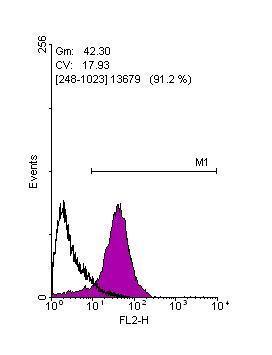
FACS, ICC, IF, IHC-Fr, IHC-P, IP, WB
Human, Mouse, Rat
Rabbit
Polyclonal
100 μl -
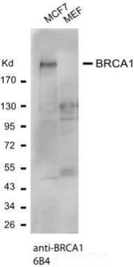
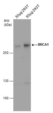
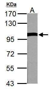
![BRCA1 antibody [17F8] - ChIP grade detects BRCA1 protein by western blot analysis. Various whole cell extracts (30 μg) were separated by 5% SDS-PAGE, and blotted with BRCA1 antibody [17F8] - ChIP grade (GRP537) diluted by 1:500. The HRP-conjugated anti](https://www.grp-ak.de/media/catalog/product/b/r/brca1-antibody-17f8-chip-grade_grp537_wb_1_2.jpg)
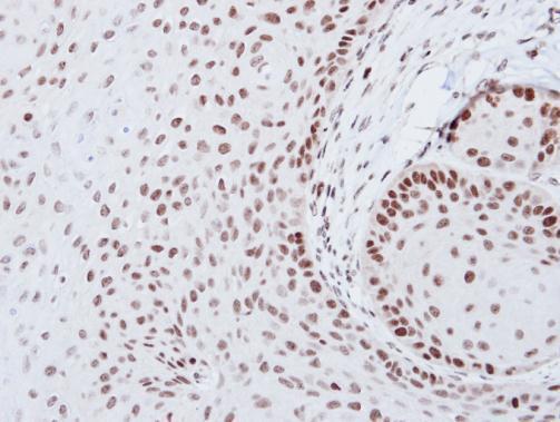
![The IHC-P analysis of BRCA1 antibody [17F8] - ChIP grade was published by Bernard-Gallon DJ and colleagues in the journal Breast Cancer Res in 2001.PMID: 11250747](https://www.grp-ak.de/media/catalog/product/b/r/brca1-antibody-17f8-chip-grade_grp537_ihc-p_2_2.jpg)
![The IHC-P analysis of BRCA1 antibody [17F8] - ChIP grade was published by Bernard-Gallon DJ and colleagues in the journal Breast Cancer Res in 2001.PMID: 11250747](https://www.grp-ak.de/media/catalog/product/b/r/brca1-antibody-17f8-chip-grade_grp537_ihc-p_1_2.jpg)
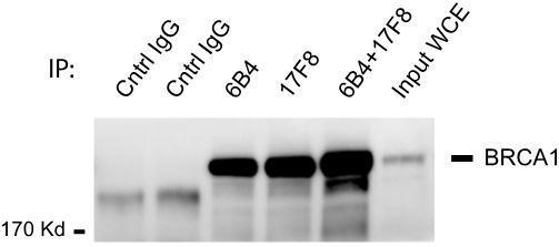

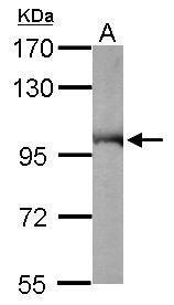
![BRCA1 antibody [6B4] (GRP538) was used at 1:1000 dilution for western blot assay of lysates from cells transfected with control or BRCA1-specific siRNA. Lysates were prepared at the indicated times following transfection. RAD50 antibody [13B3] (GRP538) wa](https://www.grp-ak.de/media/catalog/product/b/r/brca1-antibody-6b4-chip-grade_grp538_wb_2_2.jpg)
![Whole cell extract (30 μg) was separated by 7.5% SDS-PAGE, and the membrane was blotted with Mre11 antibody [12D7] (GRP540) diluted at 1:500. The HRP-conjugated anti-mouse IgG antibody was used to detect the primary antibody, and the signal was develo](https://www.grp-ak.de/media/catalog/product/m/r/mre11-antibody-12d7_grp540_wb_4_2.jpg)
![Mre11 antibody [12D7] detects Mre11 protein by western blot analysis.A. 30 μg 293T whole cell extract B. 30 μg whole cell extract of human Mre11-transfected 293T cells7.5% SDS-PAGEMre11 antibody [12D7] (GRP540) dilution: 1:1000The HRP-conjugated ant](https://www.grp-ak.de/media/catalog/product/m/r/mre11-antibody-12d7_grp540_wb_3_2.jpg)
![Various whole cell extracts (30 μg) were separated by 7.5% SDS-PAGE, and the membrane was blotted with Mre11 antibody [12D7] (GRP540) diluted at 1:1000. The HRP-conjugated anti-mouse IgG antibody was used to detect the primary antibody.](https://www.grp-ak.de/media/catalog/product/m/r/mre11-antibody-12d7_grp540_wb_2_2.jpg)
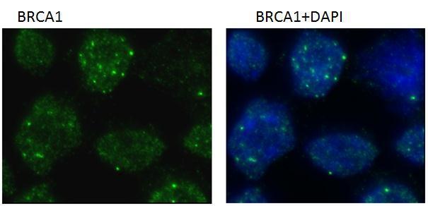
![Mre11 antibody [12D7] detects Mre11 protein at nucleus by immunofluorescent analysis.Sample: HeLa cells were fixed in 4% paraformaldehyde at RT for 15 min.Green: Mre11 stained by Mre11 antibody [12D7] (GRP540) diluted at 1:200.Blue: Hoechst 33342 staining](https://www.grp-ak.de/media/catalog/product/m/r/mre11-antibody-12d7_grp540_icc_1_2.jpg)
![The WB analysis of Mre11 antibody [12D7] was published by Harten SK and colleagues in the journal BMC Biol in 2015.PMID: 25857663](https://www.grp-ak.de/media/catalog/product/m/r/mre11-antibody-12d7_grp540_wb_1_2.jpg)
![Rad50 antibody [13B3] detects Rad50 protein at nucleus by immunofluorescent analysis.Sample: HeLa cells were fixed in 4% paraformaldehyde at RT for 15 min.Green: Rad50 protein stained by Rad50 antibody [13B3] (GRP541) diluted at 1:200.Red: phalloidin, a c](https://www.grp-ak.de/media/catalog/product/r/a/rad50-antibody-13b3_grp541_if_1_2.jpg)
![HeLa whole cell and nuclear extracts (30 μg) were separated by 5% SDS-PAGE, and the membrane was blotted with Rad50 antibody [13B3] (GRP541) diluted at 1:1000. The HRP-conjugated anti-mouset IgG antibody was used to detect the primary antibody.](https://www.grp-ak.de/media/catalog/product/r/a/rad50-antibody-13b3_grp541_wb_6_2.jpg)
![Rad50 antibody [13B3] detects Rad50 protein at nucleus in CAL 27 xenograft by immunohistochemical analysis. Sample: Paraffin-embedded CAL 27 xenograft. Rad50 antibody [13B3] (GRP541) diluted at 1:200.](https://www.grp-ak.de/media/catalog/product/r/a/rad50-antibody-13b3_grp541_ihc-p_5_2.jpg)
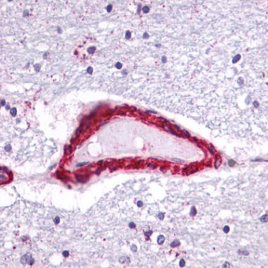
![Rad50 antibody [13B3] detects Rad50 protein at nucleus in human lung by immunohistochemical analysis. Sample: Paraffin-embedded human lung. Rad50 antibody [13B3] (GRP541) diluted at 1:200.](https://www.grp-ak.de/media/catalog/product/r/a/rad50-antibody-13b3_grp541_ihc-p_4_2.jpg)
![Rad50 antibody [13B3] detects Rad50 protein at nucleus in PC-3 xenograft by immunohistochemical analysis. Sample: Paraffin-embedded PC-3 xenograft. Rad50 antibody [13B3] (GRP541) diluted at 1:200.](https://www.grp-ak.de/media/catalog/product/r/a/rad50-antibody-13b3_grp541_ihc-p_3_2.jpg)
![Rad50 antibody [13B3] detects Rad50 protein at nucleus by immunohistochemical analysis.Sample: Paraffin-embedded human lung cancer.Rad50 stained by Rad50 antibody [13B3] (GRP541) diluted at 1:100.Antigen Retrieval: Citrate buffer, pH 6.0, 15 min](https://www.grp-ak.de/media/catalog/product/r/a/rad50-antibody-13b3_grp541_ihc-p_2_2.jpg)
![Rad50 antibody [13B3] detects Rad50 protein at nucleus by immunohistochemical analysis.Sample: Paraffin-embedded human lung cancer.Rad50 stained by Rad50 antibody [13B3] (GRP541) diluted at 1:100.Antigen Retrieval: Citrate buffer, pH 6.0, 15 min](https://www.grp-ak.de/media/catalog/product/r/a/rad50-antibody-13b3_grp541_ihc-p_1_2.jpg)
![The WB analysis of Rad50 antibody [13B3] was published by Palagyi A and colleagues in the journal Mol Cancer in 2010 .](https://www.grp-ak.de/media/catalog/product/r/a/rad50-antibody-13b3_grp541_wb_5_2.jpg)
![The WB, IP analysis of Rad50 antibody [13B3] was published by Mariggiò G and colleagues in the journal PLoS Pathog in 2017.PMID: 28430817](https://www.grp-ak.de/media/catalog/product/r/a/rad50-antibody-13b3_grp541_wb_4_2.jpg)
![The WB analysis of Rad50 antibody [13B3] was published by Mariggiò G and colleagues in the journal PLoS Pathog in 2017.PMID: 28430817](https://www.grp-ak.de/media/catalog/product/r/a/rad50-antibody-13b3_grp541_wb_3_2.jpg)
![The WB analysis of Rad50 antibody [13B3] was published by Zhu J and colleagues in the journal EMBO Mol Med in 2013.PMID: 23341130](https://www.grp-ak.de/media/catalog/product/r/a/rad50-antibody-13b3_grp541_wb_2_2.jpg)
![The WB analysis of Rad50 antibody [13B3] was published by Harten SK and colleagues in the journal BMC Biol in 2015.PMID: 25857663](https://www.grp-ak.de/media/catalog/product/r/a/rad50-antibody-13b3_grp541_wb_1_2.jpg)
![Various whole cell extracts (30 μg) were separated by 10% SDS-PAGE, and the membrane was blotted with Rad51 antibody [14B4] (GRP542) diluted at 1:500. The HRP-conjugated anti-mouset IgG antibody was used to detect the primary antibody, and the signal](https://www.grp-ak.de/media/catalog/product/r/a/rad51-antibody-14b4_grp542_wb_11_2.jpg)
![Various whole cell extracts (30 μg) were separated by 10% SDS-PAGE, and the membrane was blotted with Rad51 antibody [14B4] (GRP542) diluted at 1:500. The HRP-conjugated anti-mouset IgG antibody was used to detect the primary antibody, and the signal](https://www.grp-ak.de/media/catalog/product/r/a/rad51-antibody-14b4_grp542_wb_10_2.jpg)
![The WB analysis of Rad51 antibody [14B4] was published by Kalimutho M and colleagues in the journal Mol Oncol in 2017 .](https://www.grp-ak.de/media/catalog/product/r/a/rad51-antibody-14b4_grp542_wb_9_2.jpg)
![The WB analysis of Rad51 antibody [14B4] was published by Kalimutho M and colleagues in the journal Mol Oncol in 2017 .](https://www.grp-ak.de/media/catalog/product/r/a/rad51-antibody-14b4_grp542_wb_8_2.jpg)
![The WB analysis of Rad51 antibody [14B4] was published by Kalimutho M and colleagues in the journal Mol Oncol in 2017 .](https://www.grp-ak.de/media/catalog/product/r/a/rad51-antibody-14b4_grp542_wb_7_2.jpg)
![The WB analysis of Rad51 antibody [14B4] was published by Kalimutho M and colleagues in the journal Mol Oncol in 2017 .](https://www.grp-ak.de/media/catalog/product/r/a/rad51-antibody-14b4_grp542_wb_6_2.jpg)
![Various whole cell extracts (30 μg) were separated by 10% SDS-PAGE, and the membrane was blotted with Rad51 antibody [14B4] (GRP542) diluted at 1:500. The HRP-conjugated anti-mouse IgG antibody was used to detect the primary antibody, and the signal w](https://www.grp-ak.de/media/catalog/product/r/a/rad51-antibody-14b4_grp542_wb_5_2.jpg)
![The WB analysis of Rad51 antibody [14B4] was published by Zhu J and colleagues in the journal EMBO Mol Med in 2013.PMID: 23341130](https://www.grp-ak.de/media/catalog/product/r/a/rad51-antibody-14b4_grp542_wb_4_2.jpg)
![The WB analysis of Rad51 antibody [14B4] was published by Zhu J and colleagues in the journal EMBO Mol Med in 2013.PMID: 23341130](https://www.grp-ak.de/media/catalog/product/r/a/rad51-antibody-14b4_grp542_wb_3_2.jpg)
![The ICC/IF analysis of Rad51 antibody [14B4] was published by White MK and colleagues in the journal PLoS One in 2014.PMID: 25310191](https://www.grp-ak.de/media/catalog/product/r/a/rad51-antibody-14b4_grp542_icc_1_2.jpg)
![The WB analysis of Rad51 antibody [14B4] was published by Zhu J and colleagues in the journal EMBO Mol Med in 2013.PMID: 23341130](https://www.grp-ak.de/media/catalog/product/r/a/rad51-antibody-14b4_grp542_wb_2_2.jpg)
![Whole cell extract (30 μg) was separated by 10% SDS-PAGE, and the membrane was blotted with Rad51 antibody [14B4] (GRP542) diluted at 1:500. The HRP-conjugated anti-mouse IgG antibody was used to detect the primary antibody.](https://www.grp-ak.de/media/catalog/product/r/a/rad51-antibody-14b4_grp542_wb_1_2.jpg)
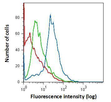
![Apolipoprotein E antibody [C2C3], C-term detects Apolipoprotein E protein at cytoplasm by immunofluorescent analysis.Sample: THP-1 cells were fixed in ice-cold MeOH for 5 min.Green: Apolipoprotein E protein stained by Apolipoprotein E antibody [C2C3], C-t](https://www.grp-ak.de/media/catalog/product/a/p/apolipoprotein-e-antibody-c2c3-c-term_grp546_if_1_2.jpg)
![Apolipoprotein E antibody [C2C3], C-term detects Apolipoprotein E protein at cytoplasm by immunofluorescent analysis.Sample: HepG2 cells were fixed in 4% paraformaldehyde at RT for 15 min.Green: Apolipoprotein E stained by Apolipoprotein E antibody [C2C3]](https://www.grp-ak.de/media/catalog/product/a/p/apolipoprotein-e-antibody-c2c3-c-term_grp546_icc_1_2.jpg)
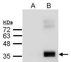
![Human plasma (30 μg) was separated by 10% SDS-PAGE, and the membrane was blotted with Apolipoprotein E antibody [C2C3], C-term (GRP546) diluted at 1:10000. The HRP-conjugated anti-rabbit IgG antibody was used to detect the primary antibody.](https://www.grp-ak.de/media/catalog/product/a/p/apolipoprotein-e-antibody-c2c3-c-term_grp546_wb_3_2.jpg)
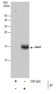
![Apolipoprotein E antibody [C2C3], C-term immunoprecipitates Apolipoprotein E protein in IP experiments.IP Sample: HepG2 whole cell lysate/extractA : 30 ?g whole cell lysate/extract of Apolipoprotein E protein expressing HepG2 cellsB : Control with 3 ?g of](https://www.grp-ak.de/media/catalog/product/a/p/apolipoprotein-e-antibody-c2c3-c-term_grp546_ip_1_2.jpg)
![Apolipoprotein E antibody [C2C3], C-term detects APOE protein by western blot analysis.A. 30 μg HepG2 whole cell lysate/extract12% SDS-PAGEApolipoprotein E antibody [C2C3], C-term (GRP546) dilution: 1:1000 The HRP-conjugated anti-rabbit IgG antibody w](https://www.grp-ak.de/media/catalog/product/a/p/apolipoprotein-e-antibody-c2c3-c-term_grp546_wb_2_2.jpg)
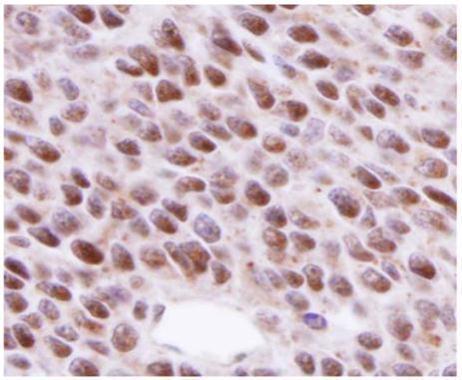
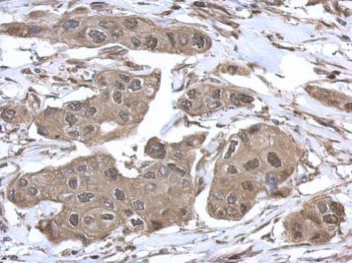
![Apolipoprotein E antibody [C2C3], C-term detects secreted Apolipoprotein E protein by immunohistochemical analysis.Sample: Paraffin-embedded human ovarian cancer.Apolipoprotein E stained by Apolipoprotein E antibody [C2C3], C-term (GRP546) diluted at 1:50](https://www.grp-ak.de/media/catalog/product/a/p/apolipoprotein-e-antibody-c2c3-c-term_grp546_ihc-p_1_2.jpg)
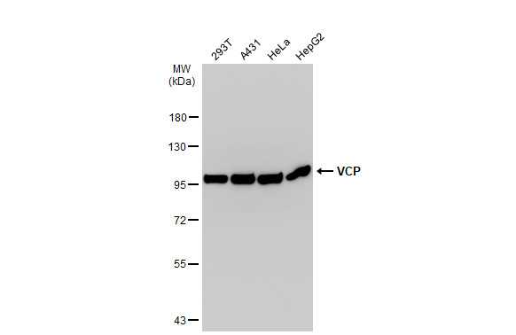
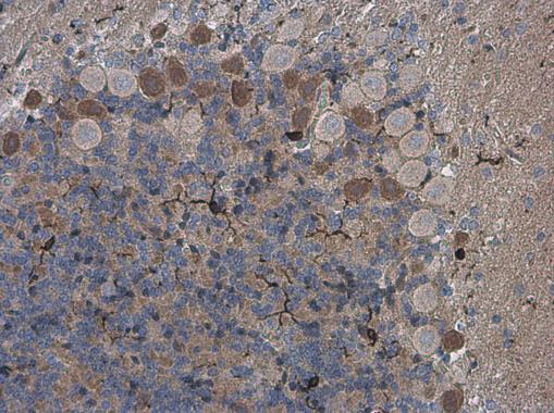
![VCP antibody detects VCP Protein expression by immunohistochemical analysis.Sample: Frozen-sectioned adult mouse cerebellum. Green: VCP stained by VCP antibody (GRP552) diluted at 1:250.Red: NF-H, stained by NF-H antibody [GT114] (GRP552) diluted at 1:500](https://www.grp-ak.de/media/catalog/product/v/c/vcp-antibody_grp552_ihc_2_2.jpg)
![C3 antibody [C3], C-term detects C3 protein at cytoplasm in mouse brain by immunohistochemical analysis. Sample: Paraffin-embedded mouse brain. C3 antibody [C3], C-term (GRP554) diluted at 1:500.](https://www.grp-ak.de/media/catalog/product/c/3/c3-antibody-c3-c-term_grp554_ihc-p_1_2.jpg)
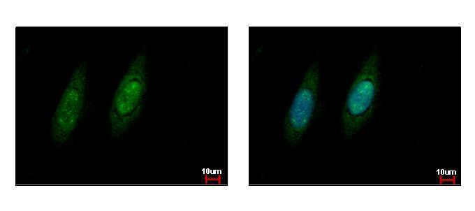
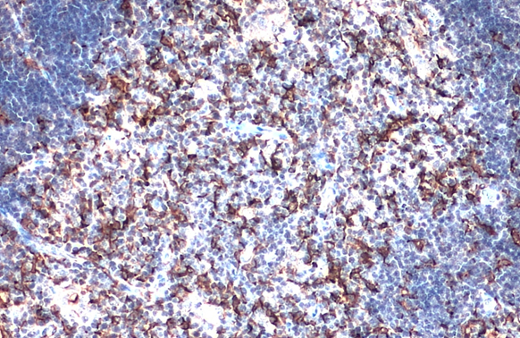
![C3 antibody [C3], C-term detects C3 protein at cytoplasm by immunofluorescent analysis.Sample: HeLa cells were fixed in 4% paraformaldehyde at RT for 15 min.Green: C3 protein stained by C3 antibody [C3], C-term (GRP554) diluted at 1:200.Blue: Hoechst 3334](https://www.grp-ak.de/media/catalog/product/c/3/c3-antibody-c3-c-term_grp554_if_1_2.jpg)

![Human plasma (30 μg) was separated by 7.5% SDS-PAGE, and the membrane was blotted with C3 antibody [C3], C-term (GRP554) diluted at 1:10000. The HRP-conjugated anti-rabbit IgG antibody was used to detect the primary antibody.](https://www.grp-ak.de/media/catalog/product/c/3/c3-antibody-c3-c-term_grp554_wb_2_2.jpg)
![Human plasma (30 μg) was separated by 7.5% SDS-PAGE, and the membrane was blotted with C3 antibody [C3], C-term (GRP554) diluted at 1:10000. The HRP-conjugated anti-rabbit IgG antibody was used to detect the primary antibody.](https://www.grp-ak.de/media/catalog/product/c/3/c3-antibody-c3-c-term_grp554_wb_1_2.jpg)
![Immunoprecipitation of C3 protein from HepG2 whole cell extracts using 5 ?g of C3 antibody [C3], C-term (GRP554).Western blot analysis was performed using C3 antibody [C3], C-term (GRP554).EasyBlot anti-Rabbit IgG was used as a secondary reagent.](https://www.grp-ak.de/media/catalog/product/c/3/c3-antibody-c3-c-term_grp554_ip_1_2.jpg)
![SOX2 antibody [N1C3] detects SOX2 protein at nucleus on rat brain stem by immunohistochemical analysis. Sample: Paraffin-embedded rat brain stem. SOX2 antibody [N1C3] (GRP557) dilution: 1:500.](https://www.grp-ak.de/media/catalog/product/s/o/sox2-antibody-n1c3_grp557_ihc_6_2.jpg)
![SOX2 antibody [N1C3] detects SOX2 protein at nucleus on mouse fore brain by immunohistochemical analysis. Sample: Paraffin-embedded mouse fore brain. SOX2 antibody [N1C3] (GRP557) diluted at 1:500.](https://www.grp-ak.de/media/catalog/product/s/o/sox2-antibody-n1c3_grp557_ihc_5_2.jpg)
![SOX2 antibody [N1C3] detects SOX2 protein at nucleus in human esophageal carcinoma by immunohistochemical analysis. Sample: Paraffin-embedded human esophageal carcinoma. SOX2 antibody [N1C3] (GRP557) diluted at 1:500.](https://www.grp-ak.de/media/catalog/product/s/o/sox2-antibody-n1c3_grp557_ihc-p_3_2.jpg)
![SOX2 antibody [N1C3] detects SOX2 protein at nucleus in human cervical carcinoma by immunohistochemical analysis. Sample: Paraffin-embedded human cervical carcinoma. SOX2 antibody [N1C3] (GRP557) diluted at 1:500.](https://www.grp-ak.de/media/catalog/product/s/o/sox2-antibody-n1c3_grp557_ihc-p_2_2.jpg)
![SOX2 antibody [N1C3] detects SOX2 protein at nucleus in mouse esophagus by immunohistochemical analysis. Sample: Paraffin-embedded mouse esophagus. SOX2 antibody [N1C3] (GRP557) diluted at 1:500.](https://www.grp-ak.de/media/catalog/product/s/o/sox2-antibody-n1c3_grp557_ihc-p_1_2.jpg)
![SOX2 antibody [N1C3] detects SOX2 protein expression by immunohistochemical analysis.Sample: Frozen-sectioned adult mouse hippocampus. Green: SOX2 protein stained by SOX2 antibody [N1C3] (GRP557) diluted at 1:250.Blue: Fluoroshield with DAPI.](https://www.grp-ak.de/media/catalog/product/s/o/sox2-antibody-n1c3_grp557_ihc_4_2.jpg)
![SOX2 antibody [N1C3] detects SOX2 protein at nucleus by immunohistochemical analysis.Sample: Frozen sectioned adult mouse retina. Green: SOX2 protein stained by SOX2 antibody [N1C3] (GRP557) diluted at 1:250.Red: Protein kinase C alpha staining.Blue: Fluo](https://www.grp-ak.de/media/catalog/product/s/o/sox2-antibody-n1c3_grp557_ihc_2_2.jpg)
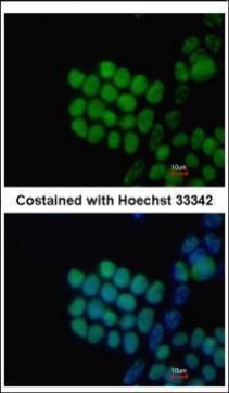
![SOX2 antibody [N1C3] detects SOX2 protein at nucleus by immunofluorescent analysis.Sample: NT2D1 cells were fixed in 4% paraformaldehyde at RT for 15 min.Green: SOX2 stained by SOX2 antibody [N1C3] (GRP557) diluted at 1:500.Red: phalloidin, a cytoskeleton](https://www.grp-ak.de/media/catalog/product/s/o/sox2-antibody-n1c3_grp557_icc_2_2.jpg)
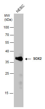
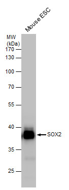
![Whole cell extract (30 μg) was separated by 12% SDS-PAGE, and the membrane was blotted with SOX2 antibody [N1C3] (GRP557) diluted at 1:10000. The HRP-conjugated anti-rabbit IgG antibody was used to detect the primary antibody.](https://www.grp-ak.de/media/catalog/product/s/o/sox2-antibody-n1c3_grp557_wb_2_2.jpg)
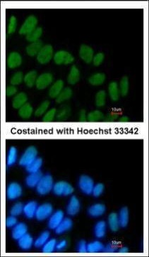
![The ICC/IF analysis of SOX2 antibody [N1C3] was published by Chang WF and colleagues in the journal PLoS One in 2016.PMID: 27802323](https://www.grp-ak.de/media/catalog/product/s/o/sox2-antibody-n1c3_grp557_icc_1_2.jpg)
![The WB analysis of SOX2 antibody [N1C3] was published by Misuno K and colleagues in the journal Stem Cell Res Ther in 2013.PMID: 24423398](https://www.grp-ak.de/media/catalog/product/s/o/sox2-antibody-n1c3_grp557_wb_1_2.jpg)
![Immunoprecipitation of SOX2 protein from NT2D1 whole cell extracts using 5 ?g of SOX2 antibody [N1C3] (GRP557).Western blot analysis was performed using SOX2 antibody [N1C3] (GRP557).EasyBlot anti-Rabbit IgG was used as a secondary reagent.](https://www.grp-ak.de/media/catalog/product/s/o/sox2-antibody-n1c3_grp557_ip_2_2.jpg)
![SOX2 antibody [N1C3] detects SOX2 protein at nucleus in mouse fetal brain by immunohistochemical analysis. Sample: Paraffin-embedded mouse fetal brain. Green: SOX2 antibody [N1C3] (GRP557) diluted at 1:200. The signal was developed using goat anti-rabbit](https://www.grp-ak.de/media/catalog/product/s/o/sox2-antibody-n1c3_grp557_ihc-p_4_2.jpg)
![Immunoprecipitation of SOX2 protein from NT2D1 whole cell extracts using 5 ?g of SOX2 antibody [N1C3] (GRP557) or SOX2 antibody [GT1876] (GRP557).Western blot analysis was performed using SOX2 antibody [N1C3] diluted at 1:500.EasyBlot anti-Rabbit IgG was](https://www.grp-ak.de/media/catalog/product/s/o/sox2-antibody-n1c3_grp557_ip_1_2.jpg)
![SOX2 antibody [N1C3] detects SOX2 protein at nucleus by immunohistochemical analysis.Sample: Frozen sectioned E13.5 rat brain. Green: SOX2 protein stained by SOX2 antibody [N1C3] (GRP557) diluted at 1:250.Red: beta Tubulin 3/ TUJ1, a mature neuron marker,](https://www.grp-ak.de/media/catalog/product/s/o/sox2-antibody-n1c3_grp557_ihc_3_2.jpg)
