Availability
- Request Lead Time
- In stock and ready for quick dispatch
- Usually dispatched within 5-10 working days
Product Overview
| Product Name | Iba1 antibody |
|---|---|
| Catalog Number | GRP104 |
| Species/Host | Rabbit |
| Reactivity | Human, Mouse, Rat |
| Conjugation | Unconjugated |
| Tested applications | FACS, ICC, IF, IHC-P, IP, WB |
| Immunogen | Recombinant protein encompassing a sequence within the center region of human Iba1. The exact sequence is proprietary. |
| Alternative Names | (click to expand) |
Product Properties
| Form/Appearance | Liquid: 1XPBS, 20% Glycerol (pH7). 0.025% ProClin 300 was added as a preservative. |
|---|---|
| Concentration | 0.56 mg/ml |
| Storage | Store as concentrated solution. Centrifuge briefly prior to opening vial. For short-term storage (1-2 weeks), store at 4°C. For long-term storage, aliquot and store at -20°C or below. Avoid multiple freeze-thaw cycles. |
| Note | For research use only. |
| Isotype | IgG |
| Clonality | Polyclonal |
| Purity | Purified by antigen-affinity chromatography. |
| Uniprot ID | P55008 |
| Entrez | 199 |
Product Description
This gene is induced by cytokines and interferon. Its protein product is thought to be involved in negative regulation of growth of vascular smooth muscle cells, which contributes to the anti-inflammatory response to vessel wall trauma. Three transcript variants encoding different isoforms have been found for this gene. [provided by RefSeq]
Application Notes
| Dilution Range | WB: 1:500-1:10000,ICC: 1:100-1:1000,IHC-P: 1:100-1:1000,FACS: 1:50-1:200,IP: 1:100-1:500 |
|---|
Validation Images
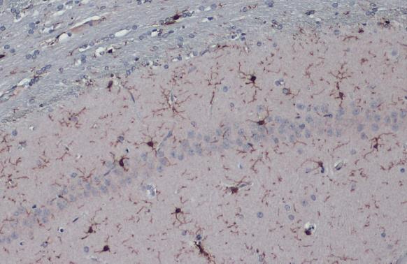
Iba1 antibody detects Iba1 protein at microglia in rat brain by immunohistochemical analysis. Sample: Paraffin-embedded rat brain. Iba1 antibody (GRP556) diluted at 1:500.
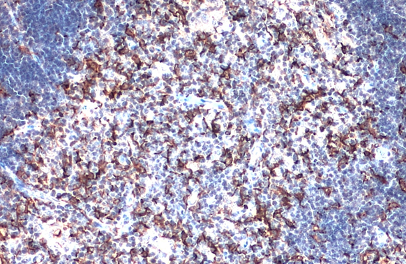
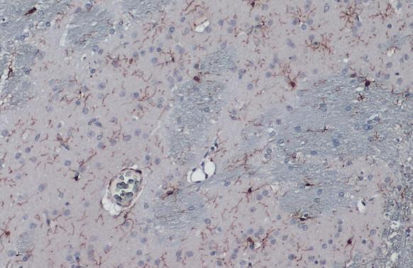
Iba1 antibody detects Iba1 protein at cytoplasm by immunohistochemical analysis.Sample: Paraffin-embedded mouse thymus gland.Iba1 stained by Iba1 antibody (GRP556) diluted at 1:500.Antigen Retrieval: Citrate buffer, pH 6.0, 15 min
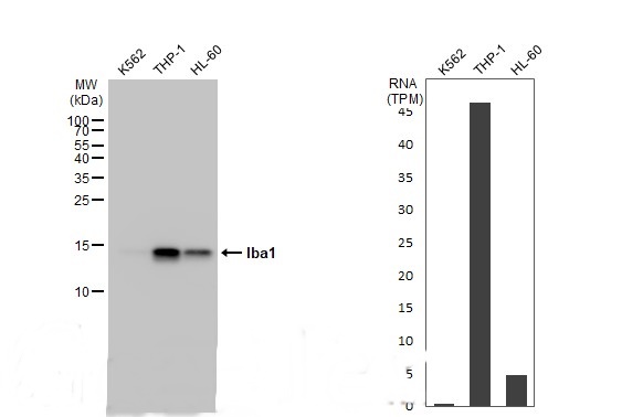
Various whole cell extracts (30 μg) were separated by 15% SDS-PAGE, and the membrane was blotted with Iba1 antibody (GRP556) diluted at 1:5000. The HRP-conjugated anti-rabbit IgG antibody was used to detect the primary antibody.Corresponding RNA expre
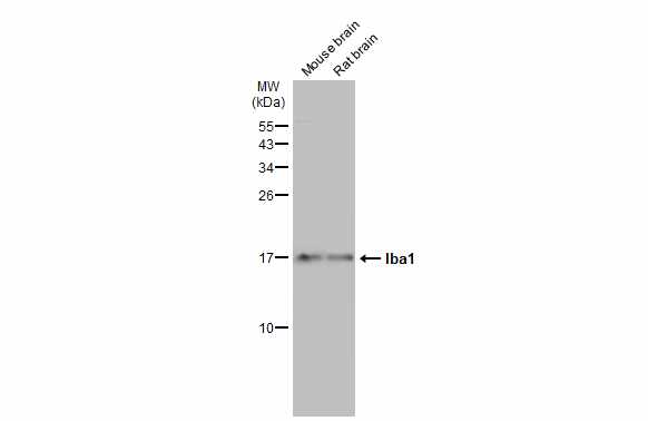
Various tissue extracts (50 μg) were separated by 15% SDS-PAGE, and the membrane was blotted with Iba1 antibody (GRP556) diluted at 1:1000. The HRP-conjugated anti-rabbit IgG antibody was used to detect the primary antibody.
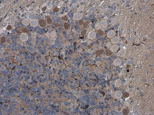
Iba1 antibody detects Iba1 protein at cytoplasm by immunohistochemical analysis.Sample: Paraffin-embedded rat brain.Iba1 stained by Iba1 antibody (GRP556) diluted at 1:2000.Antigen Retrieval: Citrate buffer, pH 6.0, 15 min
Iba1 antibody detects Iba1 protein at cytoplasm by immunohistochemical analysis.Sample: Paraffin-embedded rat brain.Iba1 stained by Iba1 antibody (GRP556) diluted at 1:2000.Antigen Retrieval: Citrate buffer, pH 6.0, 15 min
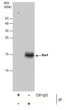
Immunoprecipitation of Iba1 protein from K562 whole cell extracts using 5 ?g of Iba1 antibody (GRP556).Western blot analysis was performed using Iba1 antibody (GRP556).EasyBlot anti-Rabbit IgG was used as a secondary reagent.
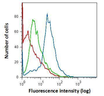
Flow cytometry on primary murine microglia cells, staining with Iba1 (GRP556) antibody using 1.0 μg per 4×10⁵ cells. (blue), Rabbit IgG (green) ,Unstained (red).
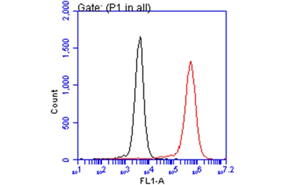
Iba1 antibody (GRP556) detects AIF1 protein by flow cytometry analysis. Sample: THP-1 cell. Black: Unlabelled sample was used as a control. Red: Iba1 antibody (GRP556) dilution: 1:50. Acquisition of 20,000 events were collected using a Dylight 488-co
Reviews
Write Your Own Review
