Primary Antibodies
- 6 images5-HT1A receptor antibody [N3C1], Internal [GRP117]
ICC, IF, IHC-Fr, IHC-P, WB
Human, Mouse, Rat
Rabbit
Polyclonal
100 μl -
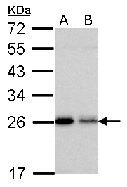
- 6 imagesMonoamine Oxidase B antibody [N2C3] [GRP118]
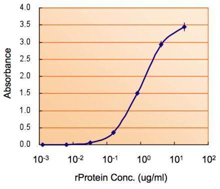
ELISA, ICC, IF, IHC-P, WB
Human, Mouse
Rabbit
Polyclonal
100 μl -
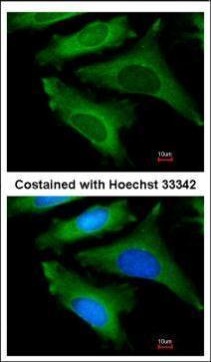
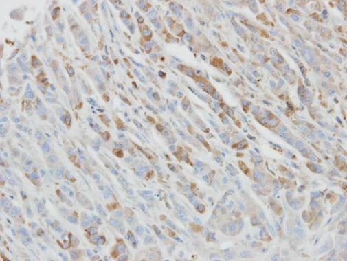
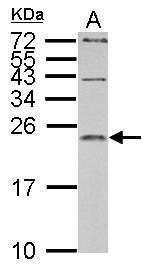
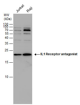
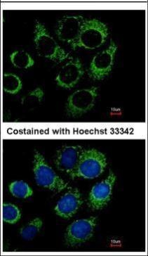
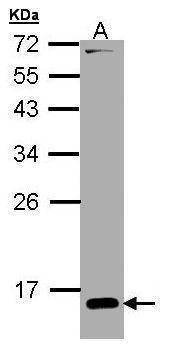
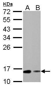
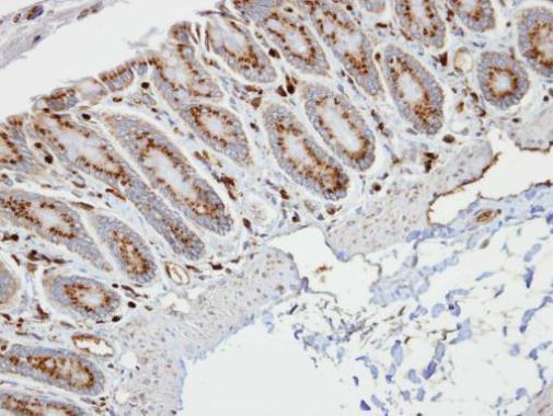
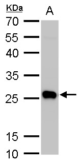
- 5 imagesIL1 Receptor antagonist antibody [GRP119]
ICC, IF, IHC-P, WB
Human, Mouse, Rat
Rabbit
Polyclonal
100 μl -
- 4 images
-
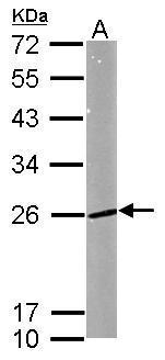
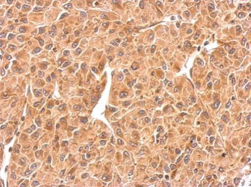
- 3 imagesMC1 Receptor antibody [C2C3], C-term [GRP123]
ICC, IF, IHC-P, WB
Human, Mouse
Rabbit
Polyclonal
100 μl -
- 13 images
-
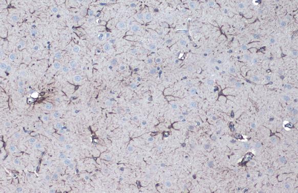
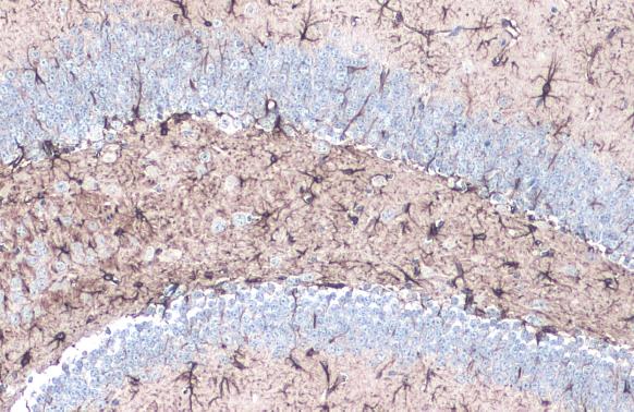
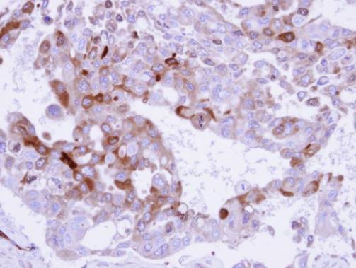
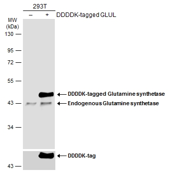
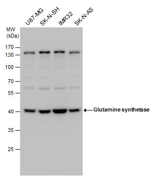
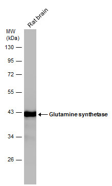
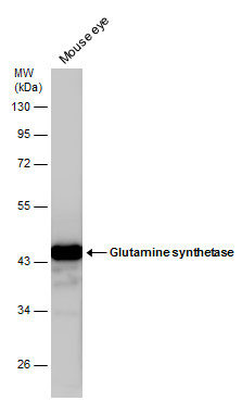
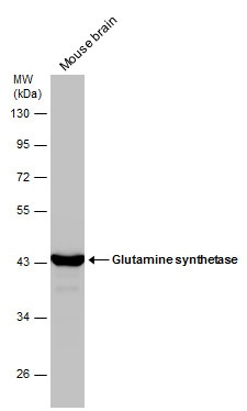
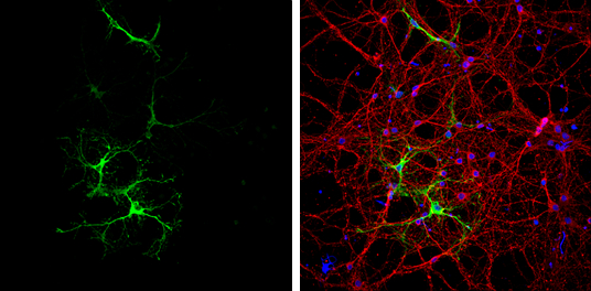
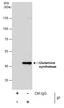
- 10 imagesGlutamine synthetase antibody [GRP125]
ICC, IF, IHC-Fr, IHC-P, IP, WB
Human, Mouse, Rat
Rabbit
Polyclonal
100 μl -
- 10 images
-
- 7 images
-
- 5 imagesALDH1A3 antibody [N2C2], Internal [GRP128]
ICC, IF, IHC-P, WB
Human, Mouse, Rat
Rabbit
Polyclonal
100 μl -
![5HT1A receptor antibody [N3C1], Internal detects 5HT1A receptor protein by western blot analysis.A. 20 μg D-hippocampus C9 lysate/extract10% SDS-PAGE5HT1A receptor antibody [N3C1], Internal (GRP569) dilution: 1:1000 The HRP-conjugated anti-rabbit IgG a](https://www.grp-ak.de/media/catalog/product/5/-/5-ht1a-receptor-antibody-n3c1-internal_grp569_wb_1_2.jpg)
![5HT1A Receptor antibody [N3C1], Internal detects 5HT1A Receptor protein at cytosol on mouse duodenum by immunohistochemical analysis. Sample: Paraffin-embedded mouse duodenum. 5HT1A Receptor antibody [N3C1], Internal (GRP569) dilution: 1:500.](https://www.grp-ak.de/media/catalog/product/5/-/5-ht1a-receptor-antibody-n3c1-internal_grp569_ihc_3_2.jpg)
![5HT1A Receptor antibody [N3C1], Internal detects 5HT1A Receptor protein at cytosol on mouse duodenum by immunohistochemical analysis. Sample: Paraffin-embedded mouse duodenum. 5HT1A Receptor antibody [N3C1], Internal (GRP569) dilution: 1:500.](https://www.grp-ak.de/media/catalog/product/5/-/5-ht1a-receptor-antibody-n3c1-internal_grp569_ihc_2_2.jpg)
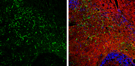
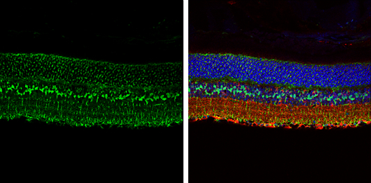
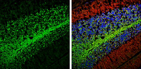
![5-HT1A receptor antibody [N3C1], Internal detects 5-HT1A receptor protein by immunohistochemical analysis. Samples: Frozen Sectioned adult mouse hippocampus.Green: 5-HT1A receptor protein stained by 5-HT1A receptor antibody [N3C1], Internal (GRP569) dilut](https://www.grp-ak.de/media/catalog/product/5/-/5-ht1a-receptor-antibody-n3c1-internal_grp569_ihc_1_2.jpg)
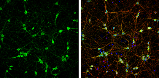
![5-HT1A receptor antibody [N3C1], Internal detects 5-HT1A receptor protein by immunofluorescent analysis.Sample: DIV14 rat E18 primary cortical neurons were fixed in 4% paraformaldehyde at RT for 15 min.Green: 5-HT1A receptor protein stained by 5-HT1A rece](https://www.grp-ak.de/media/catalog/product/5/-/5-ht1a-receptor-antibody-n3c1-internal_grp569_if_1_2.jpg)
![Monoamine Oxidase B antibody [N2C3] detects Monoamine Oxidase B protein at cytoplasm on mouse lung by immunohistochemical analysis. Sample: Paraffin-embedded mouse lung. Monoamine Oxidase B antibody [N2C3] (GRP570) diluted at 1:500.](https://www.grp-ak.de/media/catalog/product/m/o/monoamine-oxidase-b-antibody-n2c3_grp570_ihc_1_2.jpg)
![Monoamine Oxidase B antibody [N2C3] detects Monoamine Oxidase B protein at mitochondria by immunofluorescent analysis.Sample: HepG2 cells were fixed in ice-cold MeOH for 5 min.Green: Monoamine Oxidase B stained by Monoamine Oxidase B antibody [N2C3] (GRP5](https://www.grp-ak.de/media/catalog/product/m/o/monoamine-oxidase-b-antibody-n2c3_grp570_icc_1_2.jpg)
![Monoamine Oxidase B antibody [N2C3] detects Monoamine Oxidase B protein at cytoplasm by immunohistochemical analysis.Sample: Paraffin-embedded mouse liver.Monoamine Oxidase B stained by Monoamine Oxidase B antibody [N2C3] (GRP570) diluted at 1:500.Antigen](https://www.grp-ak.de/media/catalog/product/m/o/monoamine-oxidase-b-antibody-n2c3_grp570_ihc-p_1_2.jpg)
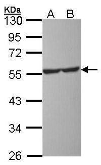
![HepG2 and mitochondria extracts (30 μg) were separated by SDS-PAGE, and the membrane was blotted with Monoamine Oxidase B antibody [N2C3] (GRP570) diluted at 1:1000. The HRP-conjugated anti-rabbit IgG antibody was used to detect the primary antibody.](https://www.grp-ak.de/media/catalog/product/m/o/monoamine-oxidase-b-antibody-n2c3_grp570_wb_1_2.jpg)
![MC1 Receptor antibody [C2C3], C-term detects MC1R protein at cytosol and membrane on human colon carcinoma by immunohistochemical analysis. Sample: Paraffin-embedded colon carcinoma. MC1 Receptor antibody [C2C3], C-term (GRP575) dilution: 1:500.](https://www.grp-ak.de/media/catalog/product/m/c/mc1-receptor-antibody-c2c3-c-term_grp575_ihc_2_2.jpg)
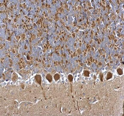
![MC1 Receptor antibody [C2C3], C-term detects MC1R protein at membrane on mouse fore brain by immunohistochemical analysis. Sample: Paraffin-embedded mouse fore brain. MC1 Receptor antibody [C2C3], C-term (GRP575) dilution: 1:500.](https://www.grp-ak.de/media/catalog/product/m/c/mc1-receptor-antibody-c2c3-c-term_grp575_ihc_1_2.jpg)
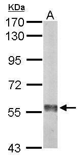
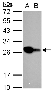
![Whole cell extract (30 μg) was separated by 10% SDS-PAGE, and the membrane was blotted with MC1 Receptor antibody [C2C3], C-term (GRP575) diluted at 1:1000. The HRP-conjugated anti-rabbit IgG antibody was used to detect the primary antibody.](https://www.grp-ak.de/media/catalog/product/m/c/mc1-receptor-antibody-c2c3-c-term_grp575_wb_1_2.jpg)
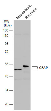
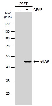
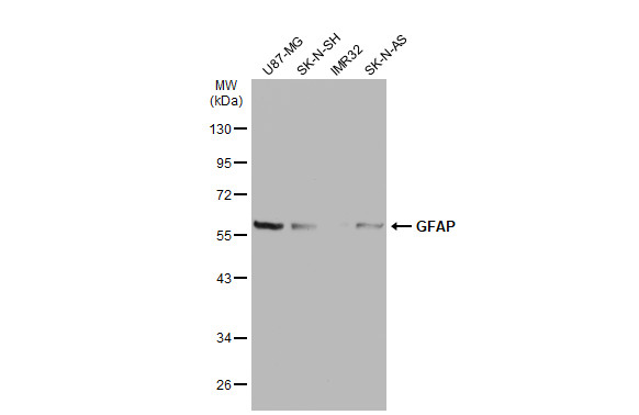
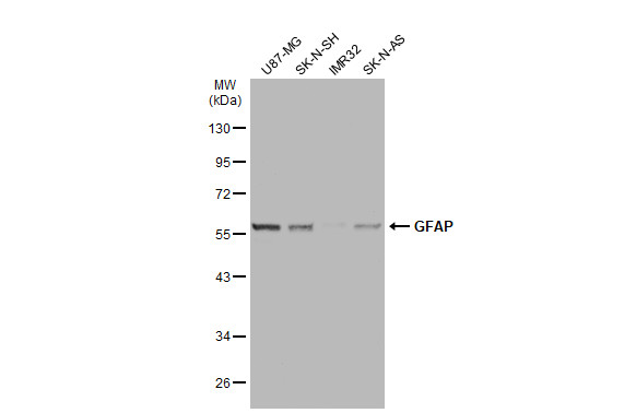
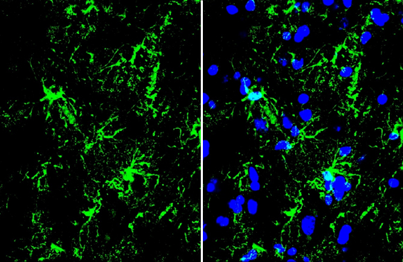
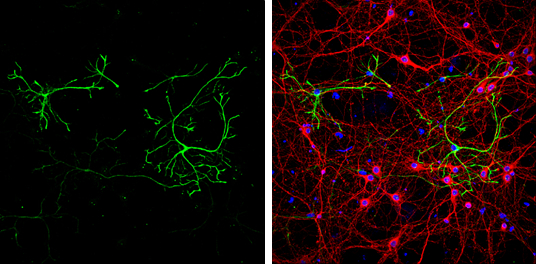
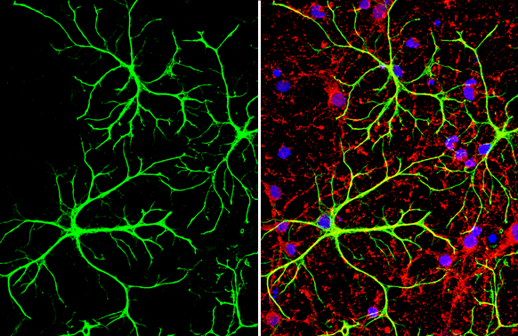
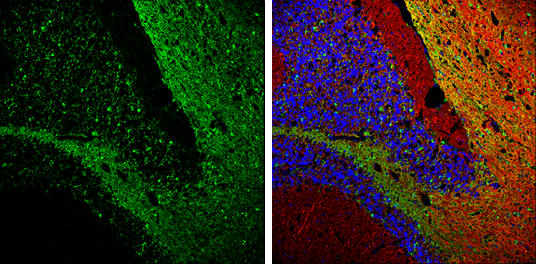
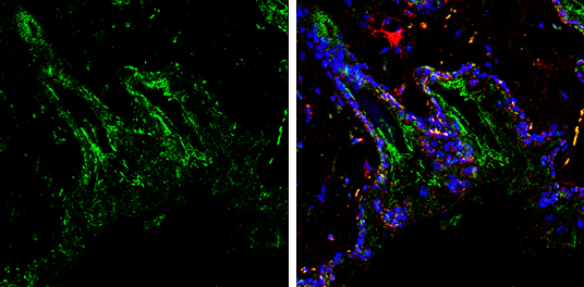
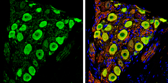
![GFAP antibody detects GFAP protein expression by immunohistochemical analysis.Sample: Frozen-sectioned adult mouse hippocampus. Green: GFAP protein stained by GFAP antibody (GRP576) diluted at 1:250.Red: NeuN, stained by NeuN antibody [2Q158] diluted at](https://www.grp-ak.de/media/catalog/product/g/f/gfap-antibody_grp576_ihc_2_2.jpg)
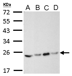
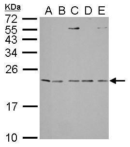
![Whole cell extract (30 μg) was separated by 10% SDS-PAGE, and the membrane was blotted with ALDH1A3 antibody [N2C2], Internal (GRP580) diluted at 1:1000. The HRP-conjugated anti-rabbit IgG antibody was used to detect the primary antibody.](https://www.grp-ak.de/media/catalog/product/a/l/aldh1a3-antibody-n2c2-internal_grp580_wb_2_2.jpg)
![Whole cell extract (30 μg) was separated by 7.5% SDS-PAGE, and the membrane was blotted with ALDH1A3 antibody [N2C2], Internal (GRP580) diluted at 1:2000.](https://www.grp-ak.de/media/catalog/product/a/l/aldh1a3-antibody-n2c2-internal_grp580_wb_1_2.jpg)
![ALDH1A3 antibody [N2C2], Internal detects ALDH1A3 protein at cytoplasm by immunofluorescent analysis.Sample: A431 cells were fixed in 4% paraformaldehyde at RT for 15 min.Green: ALDH1A3 stained by ALDH1A3 antibody [N2C2], Internal (GRP580) diluted at 1:50](https://www.grp-ak.de/media/catalog/product/a/l/aldh1a3-antibody-n2c2-internal_grp580_icc_1_2.jpg)
![ALDH1A3 antibody [N2C2], Internal detects ALDH1A3 protein at cytoplasm by immunohistochemical analysis.Sample: Paraffin-embedded rat prostate.ALDH1A3 stained by ALDH1A3 antibody [N2C2], Internal (GRP580) diluted at 1:500.Antigen Retrieval: Citrate buffer,](https://www.grp-ak.de/media/catalog/product/a/l/aldh1a3-antibody-n2c2-internal_grp580_ihc-p_2_2.jpg)
![ALDH1A3 antibody [N2C2], Internal detects ALDH1A3 protein at cytoplasm by immunohistochemical analysis.Sample: Paraffin-embedded mouse prostate.ALDH1A3 stained by ALDH1A3 antibody [N2C2], Internal (GRP580) diluted at 1:500.Antigen Retrieval: Citrate buffe](https://www.grp-ak.de/media/catalog/product/a/l/aldh1a3-antibody-n2c2-internal_grp580_ihc-p_1_2.jpg)
