Availability
- Request Lead Time
- In stock and ready for quick dispatch
- Usually dispatched within 5-10 working days
Product Overview
| Product Name | GFAP antibody |
|---|---|
| Catalog Number | GRP124 |
| Species/Host | Rabbit |
| Reactivity | Human, Mouse, Rat |
| Conjugation | Unconjugated |
| Tested applications | ICC, IF, IHC-Fr, IHC-P, WB |
| Immunogen | Recombinant protein encompassing a sequence within the center region of human GFAP. The exact sequence is proprietary. |
| Alternative Names | (click to expand) |
Product Properties
| Form/Appearance | Liquid: 1XPBS, 20% Glycerol (pH7). 0.025% ProClin 300 was added as a preservative. |
|---|---|
| Concentration | 1.13 mg/ml |
| Storage | Store as concentrated solution. Centrifuge briefly prior to opening vial. For short-term storage (1-2 weeks), store at 4°C. For long-term storage, aliquot and store at -20°C or below. Avoid multiple freeze-thaw cycles. |
| Note | For research use only. |
| Isotype | IgG |
| Clonality | Polyclonal |
| Purity | Purified by antigen-affinity chromatography. |
| Uniprot ID | P14136 |
| Entrez | 2670 |
Product Description
This gene encodes one of the major intermediate filament proteins of mature astrocytes. It is used as a marker to distinguish astrocytes from other glial cells during development. Mutations in this gene cause Alexander disease, a rare disorder of astrocytes in the central nervous system. Alternative splicing results in multiple transcript variants encoding distinct isoforms. [provided by RefSeq]
Application Notes
| Dilution Range | WB: 1:5000-1:50000,ICC: 1:100-1:1000,IHC-P: 1:100-1:1000,IHC-Fr: 1:100-1:1000 |
|---|
Validation Images
![GFAP antibody detects GFAP protein expression by immunohistochemical analysis.Sample: Frozen-sectioned adult mouse hippocampus. Green: GFAP protein stained by GFAP antibody (GRP576) diluted at 1:250.Red: NeuN, stained by NeuN antibody [2Q158] diluted at](https://www.grp-ak.de/media/catalog/product/g/f/gfap-antibody_grp576_ihc_2_2.jpg)
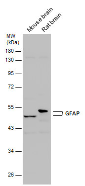
Various tissue extracts (50 μg) were separated by 10% SDS-PAGE, and the membrane was blotted with GFAP antibody (GRP576) diluted at 1:50000.
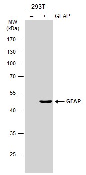
Non-transfected (–) and transfected (+) 293T whole cell extracts (30 μg) were separated by 10% SDS-PAGE, and the membrane was blotted with GFAP antibody (GRP576) diluted at 1:20000. The HRP-conjugated anti-rabbit IgG antibody was used to detect the
GFAP antibody detects GFAP protein expression by immunohistochemical analysis.Sample: Frozen-sectioned adult mouse hippocampus. Green: GFAP protein stained by GFAP antibody (GRP576) diluted at 1:250.Red: NeuN, stained by NeuN antibody [2Q158] diluted at
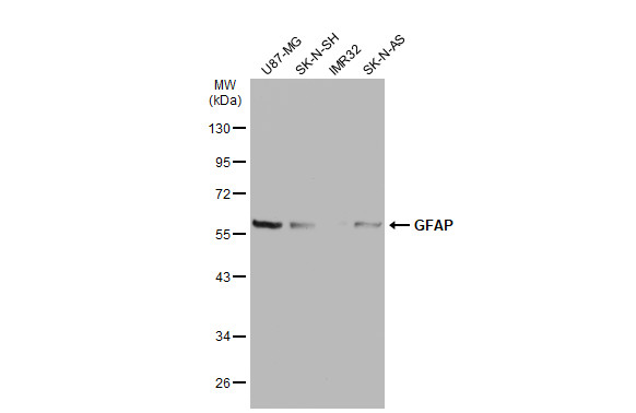
Various whole cell extracts (30 μg) were separated by 10% SDS-PAGE, and the membrane was blotted with GFAP antibody (GRP576) diluted at 1:20000. The HRP-conjugated anti-rabbit IgG antibody was used to detect the primary antibody.
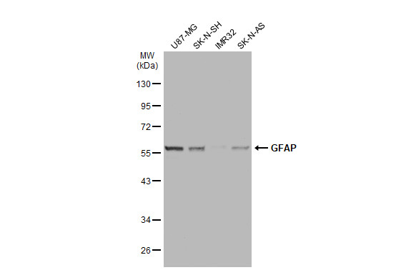
Various whole cell extracts (30 μg) were separated by 10% SDS-PAGE, and the membrane was blotted with GFAP antibody (GRP576) diluted at 1:20000. The HRP-conjugated anti-rabbit IgG antibody was used to detect the primary antibody.
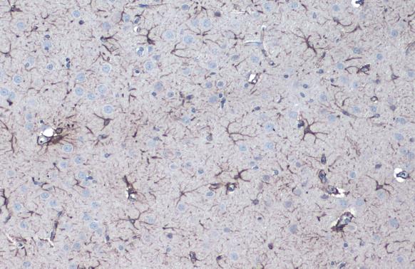
GFAP antibody detects GFAP protein at cytoplasm by immunohistochemical analysis.Sample: Paraffin-embedded rat brain.GFAP stained by GFAP antibody (GRP576) diluted at 1:1000.Antigen Retrieval: Citrate buffer, pH 6.0, 15 min
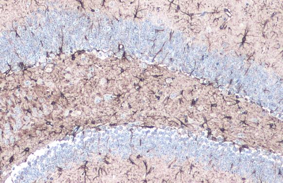
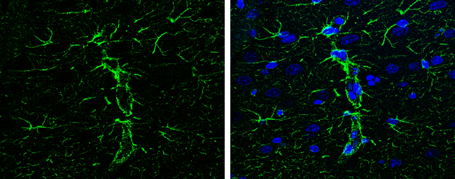
GFAP antibody detects GFAP protein at cytoplasm by immunohistochemical analysis.Sample: Paraffin-embedded mouse hippocampus.GFAP stained by GFAP antibody (GRP576) diluted at 1:1000.Antigen Retrieval: Citrate buffer, pH 6.0, 15 min
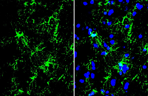
GFAP antibody detects GFAP protein by immunohistochemical analysis.Sample: Frozen-sectioned mouse spinal cord.Green: GFAP stained by GFAP antibody (GRP576) diluted at 1:300.Blue: Hoechst 33342 staining.
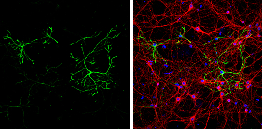
GFAP antibody detects GFAP protein at glia cells by immunofluorescent analysis.Sample: DIV9 rat E18 primary cortical neurons were fixed in 4% paraformaldehyde at RT for 15 min.Green: GFAP protein stained by GFAP antibody (GRP576) diluted at 1:500.Red: bet
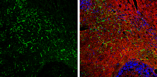
GFAP antibody detects GFAP protein expression by immunohistochemical analysis.Sample: Frozen-sectioned adult mouse cerebellum. Green: GFAP protein stained by GFAP antibody (GRP576) diluted at 1:250.Red: beta Tubulin 3/ TUJ1, stained by beta Tubulin 3/ TUJ
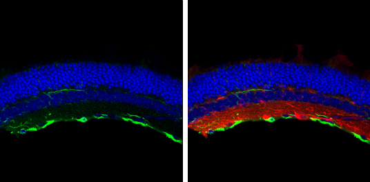
GFAP antibody detects GFAP protein at retinal ganglion cell layer by immunohistochemical analysis.Sample: Frozen sectioned adult mouse retina. Green: GFAP protein stained by GFAP antibody (GRP576) diluted at 1:250.Red: beta Tubulin 3/ TUJ1, stained by bet
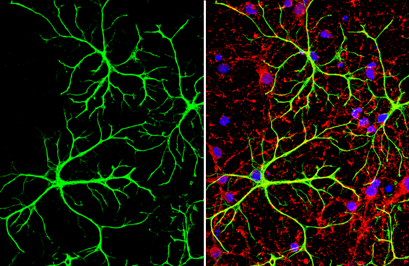
GFAP antibody detects GFAP protein at cytoplasm in rat brain by immunohistochemical analysis. Sample: Paraffin-embedded rat brain. Green: GFAP antibody (GRP576) diluted at 1:200. The signal was developed using goat anti-rabbit IgG antibody (Dylight488) (G
GFAP antibody detects GFAP protein at glia cells by immunofluorescent analysis.Sample: DIV10 rat E18 primary cortical neuron and glia cells were fixed in 4% paraformaldehyde at RT for 15 min.Green: GFAP stained by GFAP antibody (GRP576) diluted at 1:500.
Reviews
Write Your Own Review
