Availability
- Request Lead Time
- In stock and ready for quick dispatch
- Usually dispatched within 5-10 working days
Product Overview
| Product Name | Glutamine synthetase antibody |
|---|---|
| Catalog Number | GRP125 |
| Species/Host | Rabbit |
| Reactivity | Human, Mouse, Rat |
| Conjugation | Unconjugated |
| Tested applications | ICC, IF, IHC-Fr, IHC-P, IP, WB |
| Immunogen | Full length human Glutamine synthetase Recombinant protein. |
| Alternative Names | (click to expand) |
Product Properties
| Form/Appearance | Liquid: 1XPBS, 20% Glycerol (pH7). 0.025% ProClin 300 was added as a preservative. |
|---|---|
| Concentration | 1 mg/ml |
| Storage | Store as concentrated solution. Centrifuge briefly prior to opening vial. For short-term storage (1-2 weeks), store at 4°C. For long-term storage, aliquot and store at -20°C or below. Avoid multiple freeze-thaw cycles. |
| Note | For research use only. |
| Isotype | IgG |
| Clonality | Polyclonal |
| Purity | Purified by antigen-affinity chromatography. |
| Uniprot ID | P15104 |
| Entrez | 2752 |
Product Description
Glutamine is a main source of energy and is involved in cell proliferation, inhibition of apoptosis, and cell signaling (Haberle et al., 2005 [PubMed 16267323]). Fetal glutamine requirements are very high and depend largely on active glutamine synthesis and the release of glutamine into the fetal circulation by the placenta. Glutamine synthetase (EC 6.3.1.2), also called glutamate-ammonia ligase (GLUL), is expressed throughout the body and plays an important role in controlling body pH and in removing ammonia from the circulation. The enzyme clears L-glutamate, the major neurotransmitter in the central nervous system, from neuronal synapses (see references in Clancy et al., 1996 [PubMed 8975719]).[supplied by OMIM]
Application Notes
| Dilution Range | WB: 1:5000-1:20000,ICC: 1:100-1:1000,IHC-P: 1:100-1:1000,IHC-Fr: 1:100-1:1000,IP: 1:100-1:500 |
|---|
Validation Images
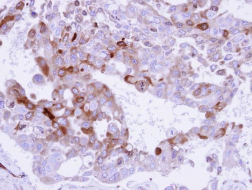
Immunohistochemical analysis of paraffin-embedded H441 xenograft , using Glutamine Synthetase (GRP577) antibody at 1:500 dilution.
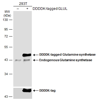
Non-transfected (–) and transfected (+) 293T whole cell extracts (30 μg) were separated by 10% SDS-PAGE, and the membrane was blotted with Glutamine synthetase antibody (GRP577) diluted at 1:10000. The HRP-conjugated anti-rabbit IgG antibody was use
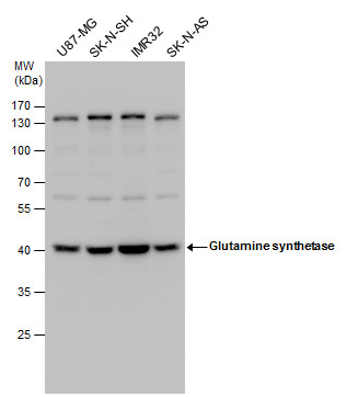
Glutamine synthetase antibody detects Glutamine synthetase protein by western blot analysis. Various whole cell extracts (30 μg) were separated by 10% SDS-PAGE, and the membrane was blotted with Glutamine synthetase antibody (GRP577) diluted at a dilut
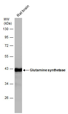
Rat tissue extract (50 μg) was separated by 10% SDS-PAGE, and the membrane was blotted with Glutamine synthetase antibody (GRP577) diluted at 1:50000.
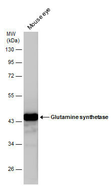
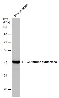
Mouse tissue extract (50 μg) was separated by 10% SDS-PAGE, and the membrane was blotted with Glutamine synthetase antibody (GRP577) diluted at 1:20000.
Mouse tissue extract (50 μg) was separated by 10% SDS-PAGE, and the membrane was blotted with Glutamine synthetase antibody (GRP577) diluted at 1:50000.
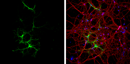
Glutamine synthetase antibody detects Glutamine synthetase protein at astrocytes by immunofluorescent analysis.Sample: DIV9 rat E18 primary cortical neurons were fixed in 4% paraformaldehyde at RT for 15 min.Green: Glutamine synthetase protein stained by
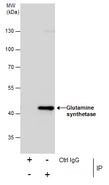
Immunoprecipitation of Glutamine synthetase protein from IMR32 whole cell extracts using 5 ?g of Glutamine synthetase antibody (GRP577).Western blot analysis was performed using Glutamine synthetase antibody (GRP577).EasyBlot anti-Rabbit IgG was used as
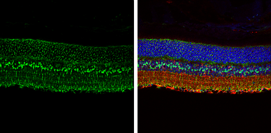
Glutamine synthetase antibody detects Glutamine synthetase protein expression by immunohistochemical analysis.Sample:Paraffin-embedded adult mouse retina. Green: Glutamine synthetase protein stained by Glutamine synthetase antibody (GRP577) diluted at 1:2
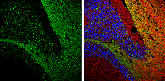
Glutamine synthetase antibody detects Glutamine synthetase protein expression by immunohistochemical analysis.Sample: Frozen-sectioned adult mouse cerebellum. Green: Glutamine synthetase protein stained by Glutamine synthetase antibody (GRP577) diluted at
Reviews
Write Your Own Review
