Search results for: 'Formyl peptide ant'
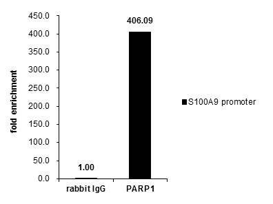
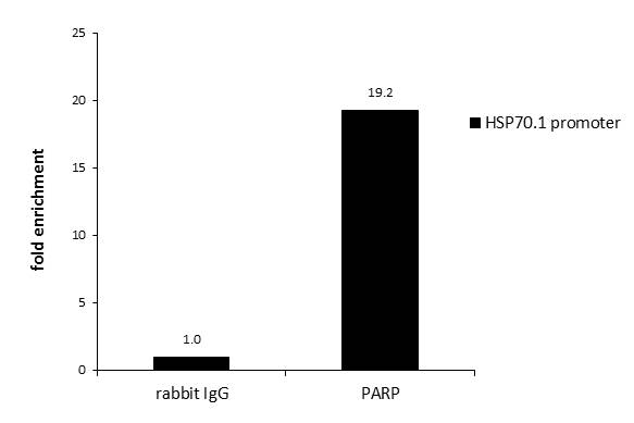
- 10 imagesPARP antibody [N2C1], Internal [GRP54]
ChIP, ICC, IF, IHC-P, IP, WB
Human, Mouse, Rat
Rabbit
Polyclonal
100 μl -
- 4 images
-
- 10 imagesAKT antibody [N3C2], Internal [GRP61]
ICC, IF, IHC-Fr, IHC-P, IP, WB
Human, Mouse, Rat, Fish
Rabbit
Polyclonal
100 μl -
- 6 images
-
- 7 imagesTET1 antibody [N3C1] [GRP63]
ChIP, ICC, IF, IHC-P, IP, WB
Human, Mouse, Monkey
Rabbit
Polyclonal
100 μl -
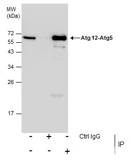
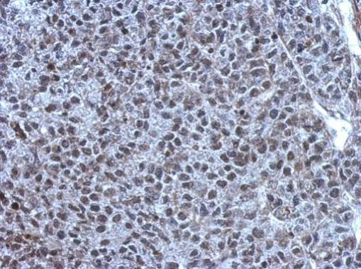
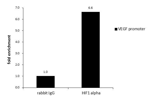
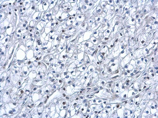
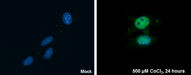
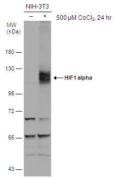
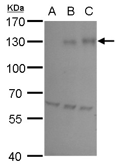
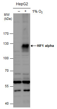
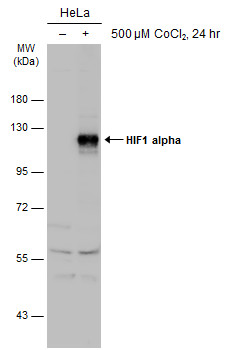
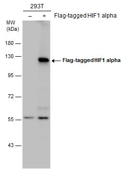
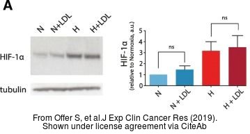
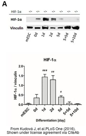
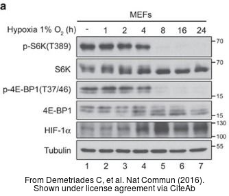
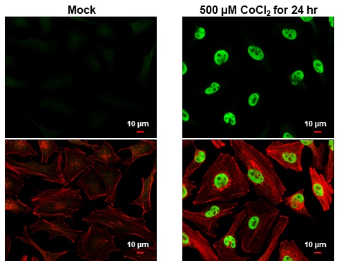
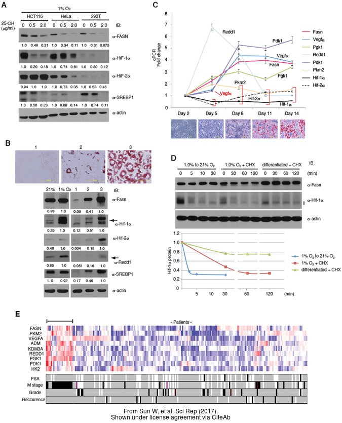
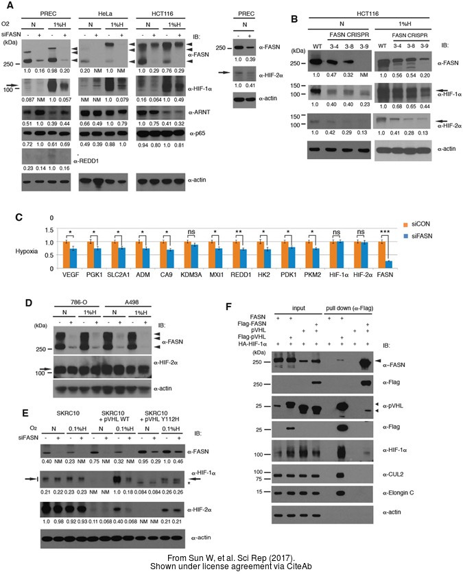
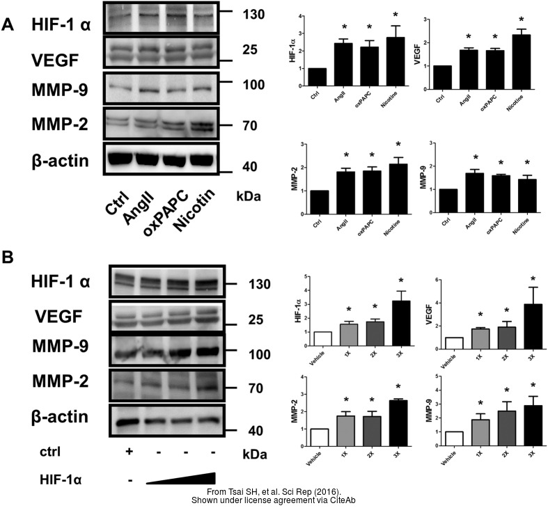
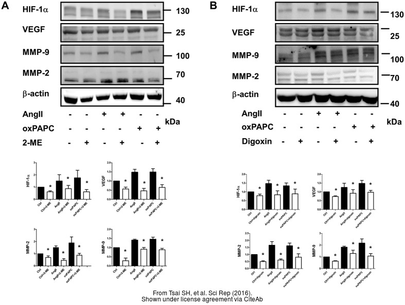
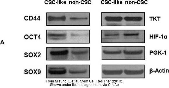
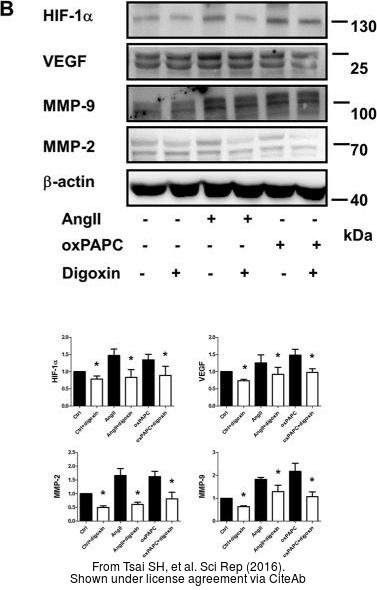
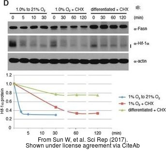
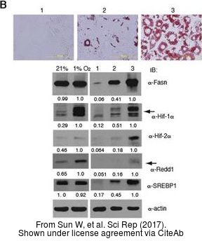
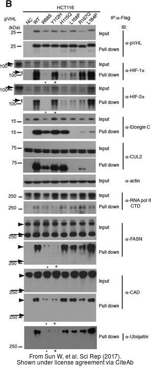
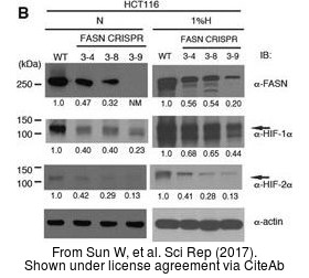
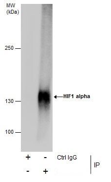
- 28 imagesHIF1 alpha antibody [GRP65]
ChIP, ICC, IF, IHC-Fr, IHC-P, IP, WB
Human, Mouse, Rat, Bovine, Rabbit
Rabbit
Polyclonal
100 μl -
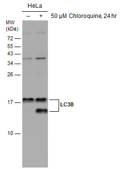
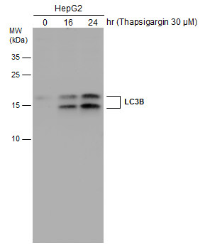
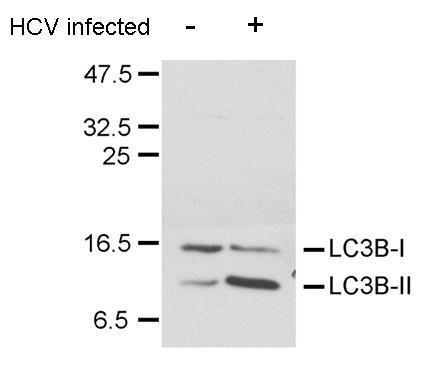
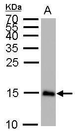
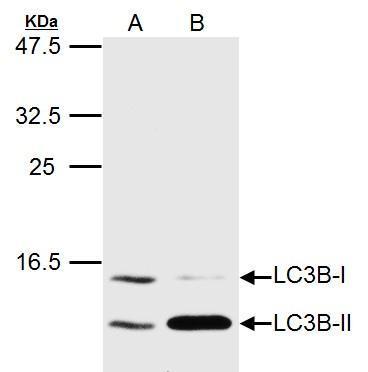
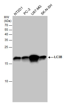
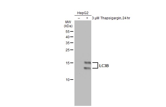
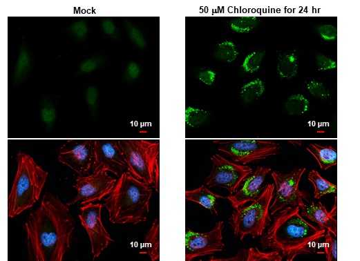
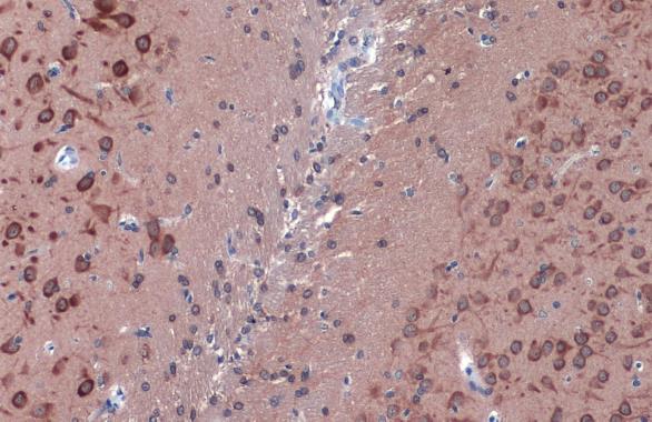
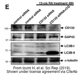
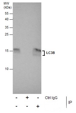
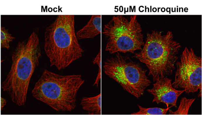
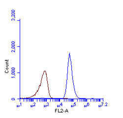
- 15 imagesLC3B antibody [GRP69]
FACS, ICC, IF, IHC-Fr, IHC-P, IP, WB
Human, Mouse, Rat, Pig
Rabbit
Polyclonal
100 μl -
- 14 images
-
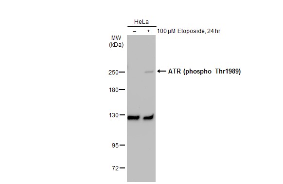
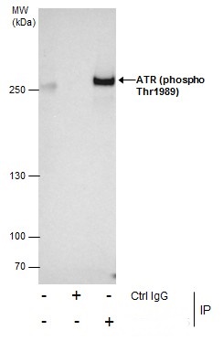
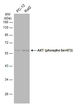
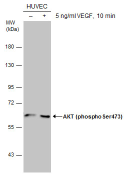
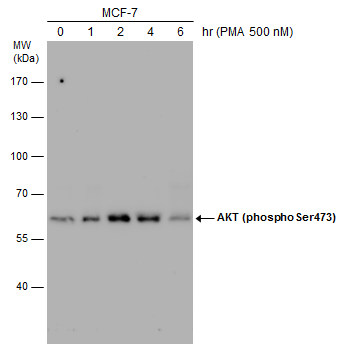
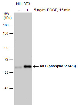
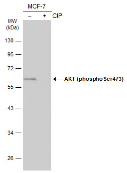
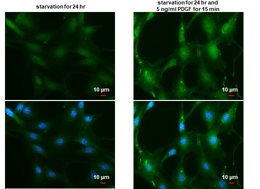
- 10 imagesAKT (phospho Ser473) antibody [GRP71]
ICC, IF, IHC-P, IP, WB
Human, Mouse, Rat
Rabbit
Polyclonal
100 μl -
- 6 images
-
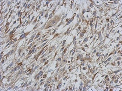
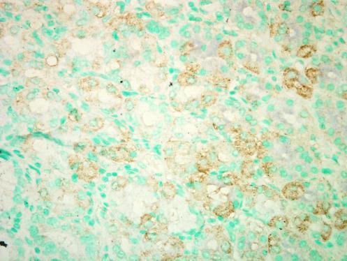
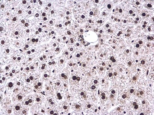
![PARP1 antibody [N2C1], Internal detects PARP1 protein at nucleus on HeLa xenograft by immunohistochemical analysis. Sample: Paraffin-embedded HeLa xenograft. PARP1 antibody [N2C1], Internal (GRP506) dilution: 1:500.](https://www.grp-ak.de/media/catalog/product/p/a/parp-antibody-n2c1-internal_grp506_ihc_1_2.jpg)
![Various whole cell extracts (30 μg) were separated by 7.5% SDS-PAGE, and the membrane was blotted with PARP1 antibody [N2C1], Internal (GRP506) diluted at 1:500. The HRP-conjugated anti-rabbit IgG antibody was used to detect the primary antibody.](https://www.grp-ak.de/media/catalog/product/p/a/parp-antibody-n2c1-internal_grp506_wb_5_2.jpg)
![Non-transfected (–) and transfected (+) 293T whole cell extracts (30 μg) were separated by 7.5% SDS-PAGE, and the membrane was blotted with PARP antibody [N2C1], Internal (GRP506) diluted at 1:50000. The HRP-conjugated anti-rabbit IgG antibody was u](https://www.grp-ak.de/media/catalog/product/p/a/parp-antibody-n2c1-internal_grp506_wb_4_2.jpg)
![Various whole cell extracts (30 μg) were separated by 5% SDS-PAGE, and the membrane was blotted with PARP antibody [N2C1], Internal (GRP506) diluted at 1:1000. The HRP-conjugated anti-rabbit IgG antibody was used to detect the primary antibody.](https://www.grp-ak.de/media/catalog/product/p/a/parp-antibody-n2c1-internal_grp506_wb_3_2.jpg)
![Untreated (–) and treated (+) HCT116 whole cell extracts (30 μg) were separated by 7.5% SDS-PAGE, and the membrane was blotted with PARP antibody [N2C1], Internal (GRP506) diluted at 1:1000. The HRP-conjugated anti-rabbit IgG antibody was used to de](https://www.grp-ak.de/media/catalog/product/p/a/parp-antibody-n2c1-internal_grp506_wb_2_2.jpg)
![Various whole cell extracts (30 μg) were separated by 5% SDS-PAGE, and the membrane was blotted with PARP antibody [N2C1], Internal (GRP506) diluted at 1:1000. The HRP-conjugated anti-rabbit IgG antibody was used to detect the primary antibody.](https://www.grp-ak.de/media/catalog/product/p/a/parp-antibody-n2c1-internal_grp506_wb_1_2.jpg)
![PARP antibody [N2C1], Internal detects PARP protein at nucleus by immunofluorescent analysis.Sample: HeLa cells were fixed in 4% paraformaldehyde at RT for 15 min.Green: PARP stained by PARP antibody [N2C1], Internal (GRP506) diluted at 1:500.Red: phalloi](https://www.grp-ak.de/media/catalog/product/p/a/parp-antibody-n2c1-internal_grp506_icc_1_2.jpg)
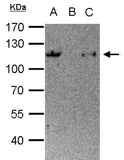
![PARP1 antibody [N2C1], Internal immunoprecipitates PARP1 protein in IP experiments.IP samples: HCT-116 whole cell extractA. 30 ?g HCT-116 whole cell extractB. Control with 4 ?g of preimmune Rabbit IgGC. Immunoprecipitation of PARP1 protein by 4 ?g PARP1 a](https://www.grp-ak.de/media/catalog/product/p/a/parp-antibody-n2c1-internal_grp506_ip_1_2.jpg)

![Non-transfected (–) and transfected (+) 293T whole cell extracts (30 μg) were separated by 10% SDS-PAGE, and the membrane was blotted with AKT antibody [N3C2], Internal (GRP513) diluted at 1:1000. The HRP-conjugated anti-rabbit IgG antibody was used](https://www.grp-ak.de/media/catalog/product/a/k/akt-antibody-n3c2-internal_grp513_wb_6_2.jpg)
![Various whole cell extracts (30 μg) were separated by 7.5% SDS-PAGE, and the membrane was blotted with AKT antibody [N3C2], Internal (GRP513) diluted at 1:1000. The HRP-conjugated anti-rabbit IgG antibody was used to detect the primary antibody, and t](https://www.grp-ak.de/media/catalog/product/a/k/akt-antibody-n3c2-internal_grp513_wb_4_2.jpg)
![AKT antibody [N3C2], Internal detects AKT protein at cytoplasm by immunofluorescent analysis.Sample: HeLa cells were fixed in 4% paraformaldehyde at RT for 15 min.Green: AKT stained by AKT antibody [N3C2], Internal (GRP513) diluted at 1:500.Blue: Hoechst](https://www.grp-ak.de/media/catalog/product/a/k/akt-antibody-n3c2-internal_grp513_icc_1_2.jpg)
![Various whole cell extracts (30 μg) were separated by 10% SDS-PAGE, and the membrane was blotted with AKT antibody [N3C2], Internal (GRP513) diluted at 1:1000. The HRP-conjugated anti-rabbit IgG antibody was used to detect the primary antibody.](https://www.grp-ak.de/media/catalog/product/a/k/akt-antibody-n3c2-internal_grp513_wb_3_2.jpg)
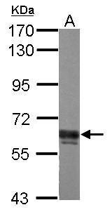
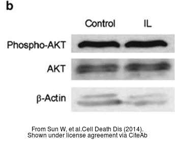
![The WB analysis of AKT antibody [N3C2], Internal was published by Sun W and colleagues in the journal Cell Death Dis in 2014 .](https://www.grp-ak.de/media/catalog/product/a/k/akt-antibody-n3c2-internal_grp513_wb_2_2.jpg)
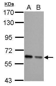
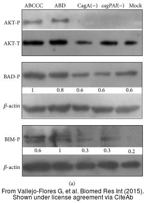
![The WB analysis of AKT antibody [N3C2], Internal was published by Vallejo-Flores G and colleagues in the journal Biomed Res Int in 2015.PMID: 26557697](https://www.grp-ak.de/media/catalog/product/a/k/akt-antibody-n3c2-internal_grp513_wb_1_2.jpg)
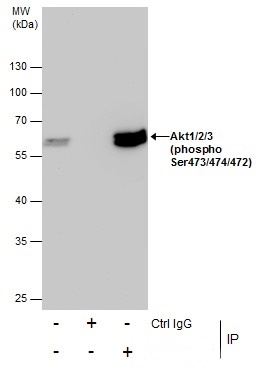
![Immunoprecipitation of Akt1/2/3 protein from 293T whole cell extracts using 5 ?g of Akt1/2/3 antibody [N3C2], Internal (GRP513).Western blot analysis was performed using Akt1/2/3 antibody [N3C2], Internal (GRP513).EasyBlot anti-Rabbit IgG was used as a s](https://www.grp-ak.de/media/catalog/product/a/k/akt-antibody-n3c2-internal_grp513_ip_1_2.jpg)

![Various whole cell extracts (30 μg) were separated by 5% SDS-PAGE, and the membrane was blotted with TET1 antibody [N3C1] (GRP515) diluted at 1:2000. The HRP-conjugated anti-rabbit IgG antibody was used to detect the primary antibody.](https://www.grp-ak.de/media/catalog/product/t/e/tet1-antibody-n3c1_grp515_wb_3_2.jpg)
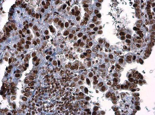
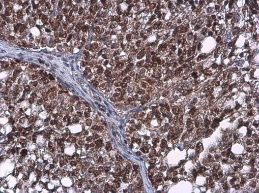
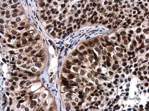
![TET1 antibody [N3C1] detects TET1 protein at nucleus in human A549 xenograft by immunohistochemical analysis. Sample: Paraffin-embedded human A549 xenograft . TET1 antibody [N3C1] (GRP515) diluted at 1:250.](https://www.grp-ak.de/media/catalog/product/t/e/tet1-antibody-n3c1_grp515_ihc-p_1_2.jpg)
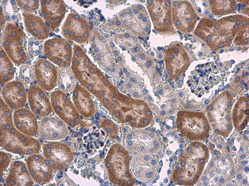
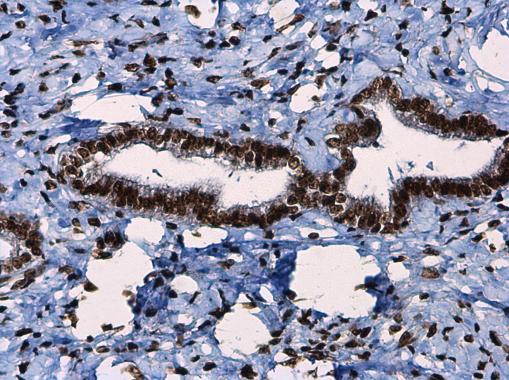
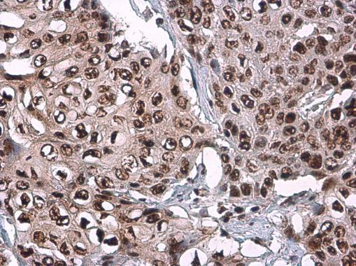
![TET1 antibody [N3C1] detects TET1 protein at nucleus on Human normal prostate tissue by immunohistochemical analysis. Sample: Paraffin-embedded Human normal prostate tissue. TET1 antibody [N3C1] (GRP515) dilution: 1:1000.](https://www.grp-ak.de/media/catalog/product/t/e/tet1-antibody-n3c1_grp515_ihc_2_2.jpg)
![HeLa whole cell and nuclear extracts (30 μg) were separated by 5% SDS-PAGE, and the membrane was blotted with TET1 antibody [N3C1] (GRP515) diluted at 1:1000. The HRP-conjugated anti-rabbit IgG antibody was used to detect the primary antibody.](https://www.grp-ak.de/media/catalog/product/t/e/tet1-antibody-n3c1_grp515_wb_2_2.jpg)
![TET1 antibody [N3C1] detects TET1 protein at nucleus by immunofluorescent analysis.Sample: Mock and transfected 293T cells were fixed in 4% paraformaldehyde at RT for 15 min.Green: TET1 stained by TET1 antibody [N3C1] (GRP515) diluted at 1:1000.Blue: Hoec](https://www.grp-ak.de/media/catalog/product/t/e/tet1-antibody-n3c1_grp515_icc_1_2.jpg)
![TET1 antibody [N3C1] detects TET1 protein by western blot analysis.A. 30 μg 293T whole cell lysate/extractB. 30 μg whole cell lysate/extract of DDDDK-human TET1-transfected 293T cells5% SDS-PAGETET1 antibody [N3C1] (GRP515) dilution: 1:5000 The HRP-](https://www.grp-ak.de/media/catalog/product/t/e/tet1-antibody-n3c1_grp515_wb_1_2.jpg)

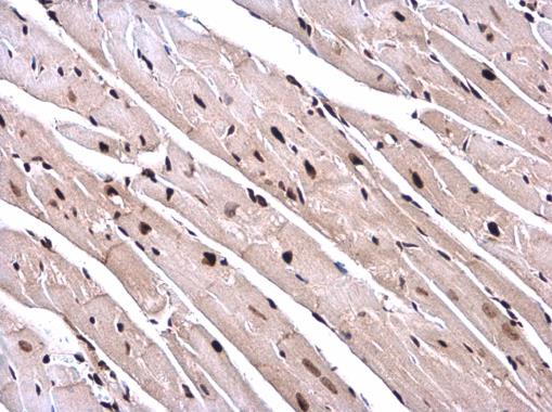
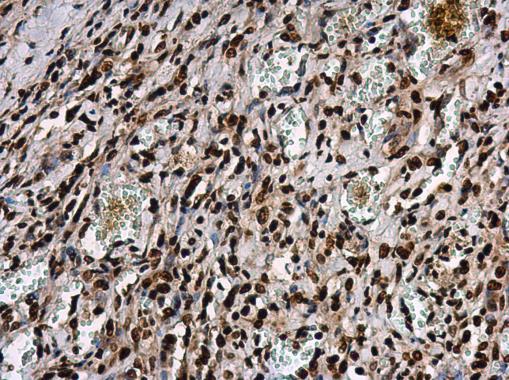
![TET1 antibody [GT1462] detects TET1 protein at nucleus on HeLa xenograft by immunohistochemical analysis. Sample: Paraffin-embedded HeLa xenograft. TET1 antibody [GT1462] (GRP530) dilution: 1:100.](https://www.grp-ak.de/media/catalog/product/t/e/tet1-antibody-gt1462_grp530_ihc_1_2.jpg)
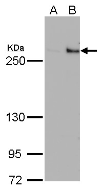
![TET1 antibody [GT1462] detects TET1 protein by western blot analysis.A. 50 μg whole cell lysate/extract from 293T cells transfected with scramble siRNA B. 50 μg whole cell lysate/extract from TET1-knockdowned 293T cells6% SDS-PAGETET1 antibody [GT14](https://www.grp-ak.de/media/catalog/product/t/e/tet1-antibody-gt1462_grp530_wb_3_2.jpg)
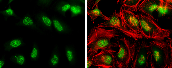
![TET1 antibody [GT1462] detects TET1 protein at nucleus by immunofluorescent analysis. Sample: TET1-transfected (right) or untransfected (left) 293T cells were fixed in 4% paraformaldehyde for 15 min. Green: TET1 protein stained by TET1 antibody (GRP530](https://www.grp-ak.de/media/catalog/product/t/e/tet1-antibody-gt1462_grp530_if_1_2.jpg)
![NT2D1 whole cell and nuclear extracts (30 μg) were separated by 5% SDS-PAGE, and the membrane was blotted with TET1 antibody [GT1462] (GRP530) diluted at 1:500.](https://www.grp-ak.de/media/catalog/product/t/e/tet1-antibody-gt1462_grp530_wb_2_2.jpg)
![Immunoprecipitation of TET1 protein from NT2D1 whole cell extracts using 5 ?g of TET1 antibody [GT1462] (GRP530).Western blot analysis was performed using TET1 antibody [GT1462] (GRP530) diluted at 1:500.EasyBlot anti-Mouse IgG was used as a secondary rea](https://www.grp-ak.de/media/catalog/product/t/e/tet1-antibody-gt1462_grp530_ip_1_2.jpg)
