Availability
- Request Lead Time
- In stock and ready for quick dispatch
- Usually dispatched within 5-10 working days
Product Overview
| Product Name | LC3B antibody |
|---|---|
| Catalog Number | GRP69 |
| Species/Host | Rabbit |
| Reactivity | Human, Mouse, Rat, Pig |
| Conjugation | Unconjugated |
| Tested applications | FACS, ICC, IF, IHC-Fr, IHC-P, IP, WB |
| Immunogen | Carrier-protein conjugated synthetic peptide encompassing a sequence within the N-terminus region of human LC3B. The exact sequence is proprietary. |
| Alternative Names | (click to expand) |
Product Properties
| Form/Appearance | Liquid: 1XPBS, 1% BSA, 20% Glycerol (pH7). 0.025% ProClin 300 was added as a preservative. |
|---|---|
| Concentration | 0.14 mg/ml |
| Storage | Store as concentrated solution. Centrifuge briefly prior to opening vial. For short-term storage (1-2 weeks), store at 4°C. For long-term storage, aliquot and store at -20°C or below. Avoid multiple freeze-thaw cycles. |
| Note | For research use only. |
| Isotype | IgG |
| Clonality | Polyclonal |
| Purity | Purified by antigen-affinity chromatography. |
| Uniprot ID | Q9GZQ8 |
| Entrez | 81631 |
Product Description
The product of this gene is a subunit of neuronal microtubule-associated MAP1A and MAP1B proteins, which are involved in microtubule assembly and important for neurogenesis. Studies on the rat homolog implicate a role for this gene in autophagy, a process that involves the bulk degradation of cytoplasmic component. [provided by RefSeq]
Application Notes
| Dilution Range | WB: 1:500-1:3000,ICC: 1:100-1:1000,IHC-P: 1:100-1:1000,FACS: 1:50-1:200,IP: 1:100-1:500 |
|---|
Validation Images
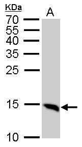
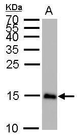
LC3B antibody detects MAP1LC3B protein by western blot analysis.A. 50 μg Rat brain lysate/extract15% SDS-PAGELC3B antibody (GRP521) dilution: 1:1000 The HRP-conjugated anti-rabbit IgG antibody was used to detect the primary antibody.
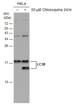
Untreated (–) and treated (+) HeLa whole cell extracts (30 μg) were separated by 15% SDS-PAGE, and the membrane was blotted with LC3B antibody (GRP521) diluted at 1:2500. The HRP-conjugated anti-rabbit IgG antibody was used to detect the primary ant
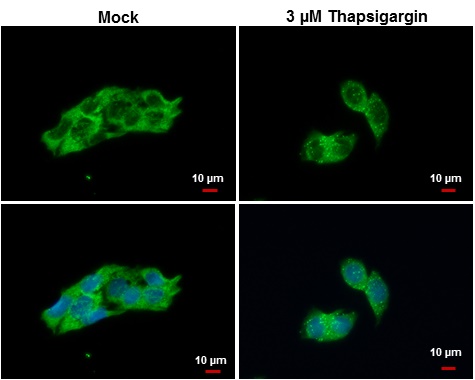
LC3B antibody detects LC3B protein at autophagosome by immunofluorescent analysis. Samples: Hep G2 cells mock (left) and treated with 3 ?M Thapsigargin for 12 hrs (right) were fixed in ice-cold MeOH for 10 min and permeabilized with ice-cold acetone for 1
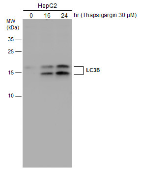
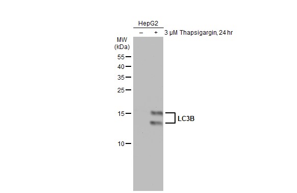
HepG2 cells were untreated or treated with 3 ?M thapsigargin for 16 and 24 hrs. Whole cell extracts (30 μg) were separated by 15% SDS-PAGE, and the membrane was blotted with LC3B antibody (GRP521) diluted at 1:1000. The HRP-conjugated anti-rabbit IgG a
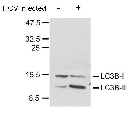
LC3B antibody detects LC3B protein in HCV-infected samples by western blot analysis. Â A. 20 μg Huh7 whole cell lysate/extract (un-infected) Â B. 20 μg Huh7 whole cell lysate/extract (HCV-infected) Â LC3B antibody (GRP521) dilution: 1:1500 Â The
LC3B antibody detects MAP1LC3B protein by western blot analysis.A. 50 μg mouse brain lysate/extract15% SDS-PAGELC3B antibody (GRP521) dilution: 1:1000 The HRP-conjugated anti-rabbit IgG antibody was used to detect the primary antibody.
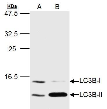
LC3B antibody detects MAP1LC3B protein by western blot analysis.A. 20 μg Huh7 whole cell lysate/extract (untreated) B. 20 μg Huh7 whole cell lysate/extract (3uM-Thapsigargin treatment for 12hr)LC3B antibody (GRP521) dilution: 1:1500 The HRP-conjugat
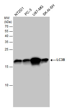
LC3B antibody detects LC3B protein by western blot analysis. Various whole cell extracts (30 μg) were separated by 15% SDS-PAGE, and the membrane was blotted with LC3B antibody (GRP521) diluted at a dilution of 1:1000. The HRP-conjugated anti-rabbit Ig
Untreated (–) and treated (+) HepG2 whole cell extracts (30 μg) were separated by 15% SDS-PAGE, and the membrane was blotted with LC3B antibody (GRP521) diluted at 1:1000. The HRP-conjugated anti-rabbit IgG antibody was used to detect the primary an
LC3B antibody detects LC3B protein at autophagosome by immunofluorescent analysis.Sample: Mock and treated HeLa cells were fixed in 4% paraformaldehyde at RT for 15 min.Green: LC3B stained by LC3B antibody (GRP521) diluted at 1:500.Red: phalloidin, a cy
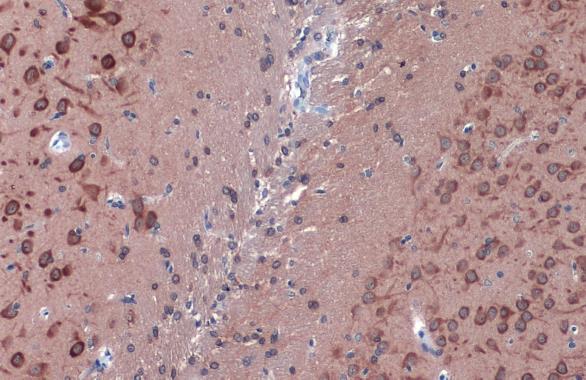
LC3B antibody detects LC3B protein at cytoplasm by immunohistochemical analysis.Sample: Paraffin-embedded rat brain.LC3B stained by LC3B antibody (GRP521) diluted at 1:500.Antigen Retrieval: Citrate buffer, pH 6.0, 15 min
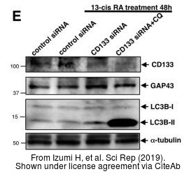
The WB analysis of LC3B antibody was published by Izumi H and colleagues in the journal Sci Rep in 2019.PMID: 30783186
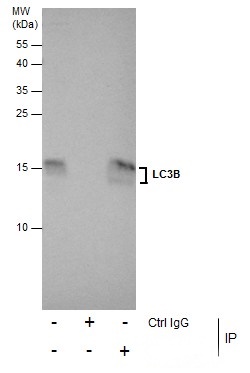
Immunoprecipitation of LC3B protein from U87-MG whole cell extracts using 5 ?g of LC3B antibody (GRP521).Western blot analysis was performed using LC3B antibody (GRP521).EasyBlot anti-Rabbit IgG was used as a secondary reagent.
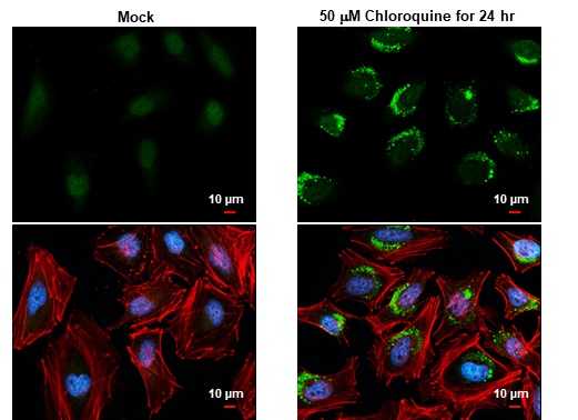
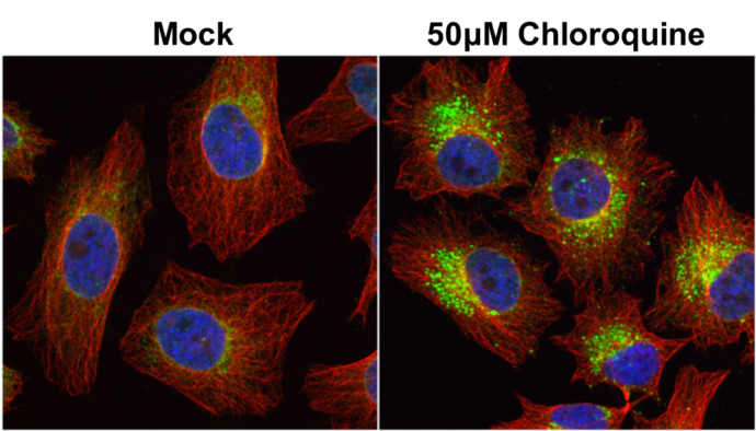
LC3B antibody detects LC3B protein at autophagosome by immunofluorescent analysis. Samples: HeLa cells mock (left) and treated with 50?M Chloroquine for 24 hr (right) were fixed in 4% paraformaldehyde at RT for 15 min.Green: LC3B protein stained by LC3B a
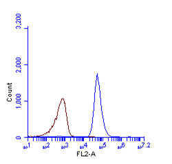
LC3B antibody (GRP521) detects LC3B protein by flow cytometry analysis. Sample: HeLa cell fixed in 4% paraformaldehyde at 4ºC for 5 min. Brown: Unlabelled sample was also used as a control. Blue: LC3B antibody (GRP521) dilution: 1:100. Acquisition of >20
Reviews
Write Your Own Review
