Search results for: 'ant'
- 2 imagesCXCR7 antibody [C1C2], Internal [GRP2]
FACS, ICC, IF, IHC-Fr, IHC-P, IP, WB
Human, Mouse
Rabbit
Polyclonal
-
- 9 imagesSQSTM1 / P62 antibody [N3C1], Internal [GRP15]
FACS, ICC, IF, IHC-P, IP, WB
Human, Mouse, Rat, Bovine, Zebrafish, Honeybee
Rabbit
Polyclonal
100 μl -
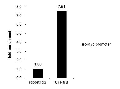
- 22 imagesbeta Catenin antibody [N1N2-2], N-term [GRP22]
ChIP, FACS, ICC, IF, IHC-P, IP, WB
Human, Mouse, Rat, Rabbit
Rabbit
Polyclonal
100 μl -
- 11 images
-
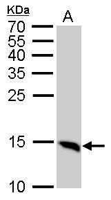
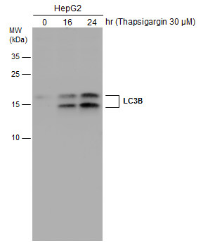
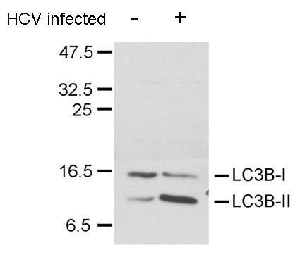
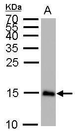
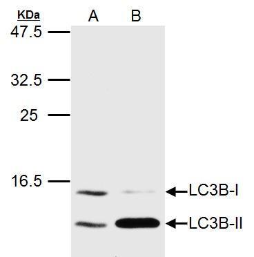
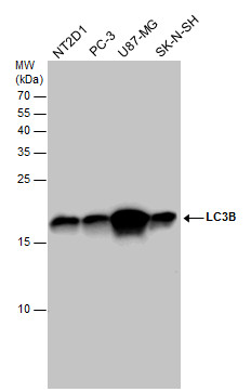
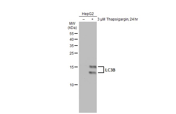
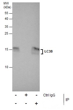
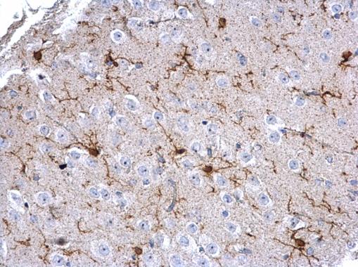
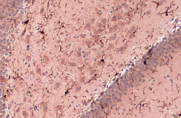
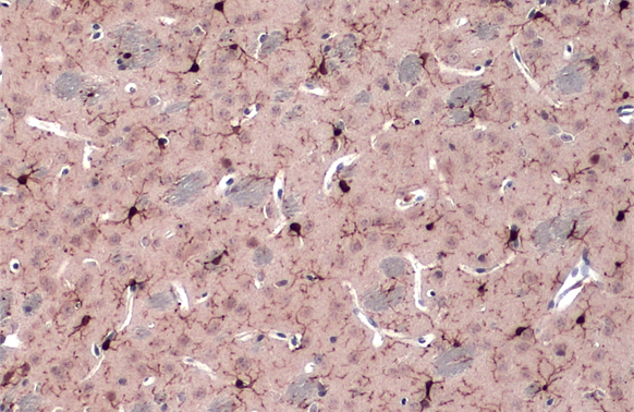
- 15 imagesLC3B antibody [GRP69]
FACS, ICC, IF, IHC-Fr, IHC-P, IP, WB
Human, Mouse, Rat, Pig
Rabbit
Polyclonal
100 μl -
- 14 images
-
- 9 images
-
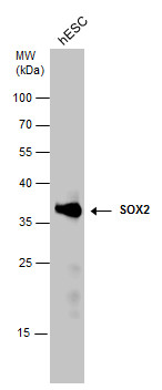
- 21 imagesSOX2 antibody [N1C3] [GRP105]
FACS, ICC, IF, IHC-Fr, IHC-P, IP, WB
Human, Mouse, Rat
Rabbit
Polyclonal
100 μl -
- 7 imagesGRK2 antibody [C2C3], C-term [GRP107]
FACS, ICC, IF, IHC-P, IP, WB
Human, Mouse
Rabbit
Polyclonal
100 μl -
- 3 images
-
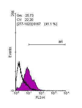
![SQSTM1 antibody [N3C1], Internal (GRP467) detects SQSTM1 protein by flow cytometry analysis.Sample: HeLa cell fixed in 4% paraformaldehyde at 4ºC for 5 min.Brown: Unlabelled sample was also used as a control.Blue: SQSTM1 antibody [N3C1], Internal] dilut](https://www.grp-ak.de/media/catalog/product/s/q/sqstm1--p62-antibody-n3c1-internal_grp467_facs_1_2.jpg)
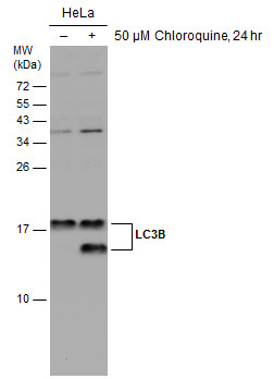
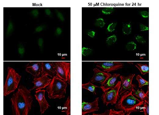
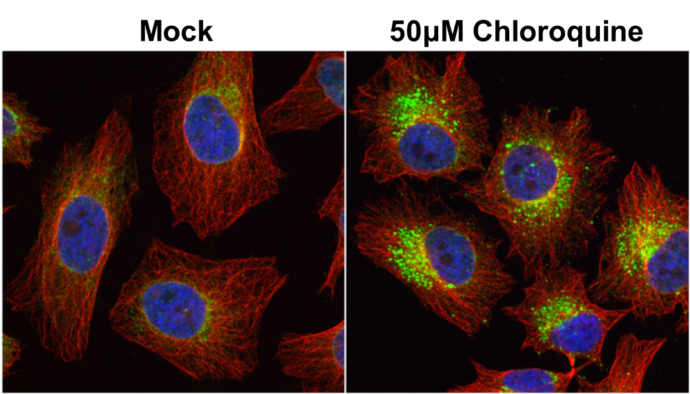
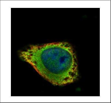
![SQSTM1 antibody [N3C1], Internal detects SQSTM1 protein at autophagosome by immunofluorescent analysis. Samples: HeLa cells mock (left) and treated with 50?M Chloroquine for 24 hr (right) were fixed in 4% paraformaldehyde at RT for 15 min.Green: SQSTM1 pr](https://www.grp-ak.de/media/catalog/product/s/q/sqstm1--p62-antibody-n3c1-internal_grp467_if_2_2.jpg)
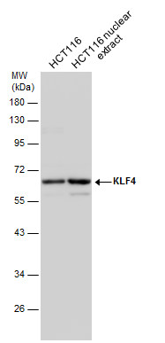
![Untreated (–) and treated (+) HepG2 whole cell extracts (30 μg) were separated by 10% SDS-PAGE, and the membrane was blotted with SQSTM1 antibody [N3C1], Internal (GRP467) diluted at 1:1000. The HRP-conjugated anti-rabbit IgG antibody was used to de](https://www.grp-ak.de/media/catalog/product/s/q/sqstm1--p62-antibody-n3c1-internal_grp467_wb_4_2.jpg)
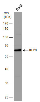
![SQSTM1 antibody [N3C1], Internal detects SQSTM1 protein by western blot analysis.A. 30 μg PC-12 whole cell lysate/extract B. 30 μg Rat2 whole cell lysate/extract10% SDS-PAGESQSTM1 antibody [N3C1], Internal (GRP467) dilution: 1:1000 The HRP-conjugate](https://www.grp-ak.de/media/catalog/product/s/q/sqstm1--p62-antibody-n3c1-internal_grp467_wb_3_2.jpg)
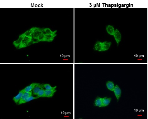
![SQSTM1 antibody [N3C1], Internal detects SQSTM1 protein at autophagosome by immunofluorescent analysis.Samples: HepG2 cells treated with 3?M thapsigargin 12 hrs (rigtht) and mock (left) were fixed in ice-cold MeOH for 10 min, permeabilize with cooled acet](https://www.grp-ak.de/media/catalog/product/s/q/sqstm1--p62-antibody-n3c1-internal_grp467_if_1_2.jpg)
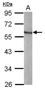
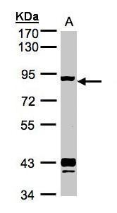
![SQSTM1 antibody [N3C1], Internal detects SQSTM1 protein by western blot analysis.A. 30 μg NIH-3T3 whole cell lysate/extract B. 30 μg JC whole cell lysate/extract C. 30 μg BCL-1 whole cell lysate/extract 12% SDS-PAGESQSTM1 antibody [N3C1], Interna](https://www.grp-ak.de/media/catalog/product/s/q/sqstm1--p62-antibody-n3c1-internal_grp467_wb_2_2.jpg)
![Untreated (–) and treated (+) Huh-7 whole cell extracts (30 μg) were separated by 10% SDS-PAGE, and the membrane was blotted with SQSTM1 antibody [N3C1], Internal (GRP467) diluted at 1:1000. The HRP-conjugated anti-rabbit IgG antibody was used to de](https://www.grp-ak.de/media/catalog/product/s/q/sqstm1--p62-antibody-n3c1-internal_grp467_wb_1_2.jpg)
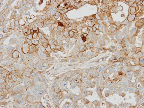
![SQSTM1 / P62 antibody [N3C1], Internal detects SQSTM1 / P62 protein at cytoplasm by immunohistochemical analysis.Sample: Paraffin-embedded human lung cancer.SQSTM1 / P62 stained by SQSTM1 / P62 antibody [N3C1], Internal (GRP467) diluted at 1:500.Antigen R](https://www.grp-ak.de/media/catalog/product/s/q/sqstm1--p62-antibody-n3c1-internal_grp467_ihc-p_1_2.jpg)
![Immunoprecipitation of SQSTM1 protein from HeLa whole cell extracts using 5 ?g of SQSTM1 antibody [N3C1], Internal (GRP467).Western blot analysis was performed using SQSTM1 antibody [N3C1], Internal (GRP467).EasyBlot anti-Rabbit IgG was used as a seconda](https://www.grp-ak.de/media/catalog/product/s/q/sqstm1--p62-antibody-n3c1-internal_grp467_ip_1_2.jpg)
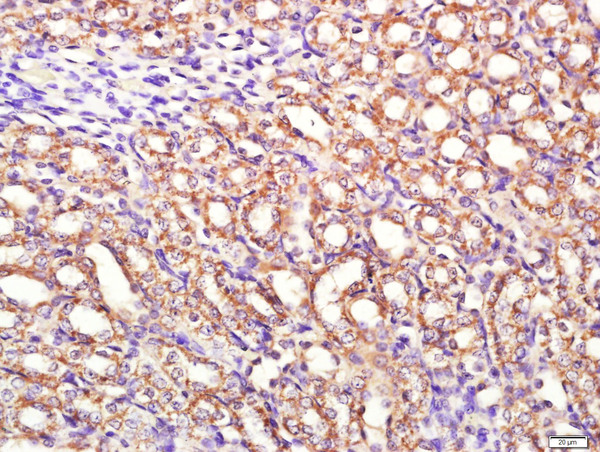
![beta Catenin antibody [N1N2-2], N-term detects beta Catenin protein at cell membrane and cytoplasm in rat colon by immunohistochemical analysis. Sample: Paraffin-embedded rat colon. beta Catenin antibody [N1N2-2], N-term (GRP474) diluted at 1:500.](https://www.grp-ak.de/media/catalog/product/b/e/beta-catenin-antibody-n1n2-2-n-term_grp474_ihc-p_9_2.jpg)
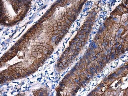
![beta Catenin antibody [N1N2-2], N-term detects beta Catenin protein at cell membrane and cytoplasm in mouse intestine by immunohistochemical analysis. Sample: Paraffin-embedded mouse intestine. beta Catenin antibody [N1N2-2], N-term (GRP474) diluted at 1:](https://www.grp-ak.de/media/catalog/product/b/e/beta-catenin-antibody-n1n2-2-n-term_grp474_ihc-p_8_2.jpg)
![beta Catenin antibody [N1N2-2], N-term detects beta Catenin protein at membrane on mouse skin by immunohistochemical analysis. Sample: Paraffin-embedded mouse skin. beta Catenin antibody [N1N2-2], N-term (GRP474) dilution: 1:500.](https://www.grp-ak.de/media/catalog/product/b/e/beta-catenin-antibody-n1n2-2-n-term_grp474_ihc_3_2.jpg)
![beta Catenin antibody [N1N2-2], N-term detects CTNNB1 protein by western blot analysis.A. 30 μg PC-12 whole cell lysate/extract](https://www.grp-ak.de/media/catalog/product/b/e/beta-catenin-antibody-n1n2-2-n-term_grp474_wb_4_2.jpg)
![7.5% SDS-PAGEbeta Catenin antibody [N1N2-2], N-term (GRP474) dilution: 1:1000 The HRP-conjugated anti-rabbit IgG antibody was used to detect the primary antibody.](https://www.grp-ak.de/media/catalog/product/b/e/beta-catenin-antibody-n1n2-2-n-term_grp474_if_3_2.jpg)
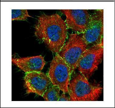
![beta Catenin antibody [N1N2-2], N-term detects beta Catenin protein at cell membrane by immunofluorescent analysis.Sample: HCT 116 cells were fixed in 4% paraformaldehyde at RT for 15 min.Green: beta Catenin protein stained by beta Catenin antibody [N1N2-](https://www.grp-ak.de/media/catalog/product/b/e/beta-catenin-antibody-n1n2-2-n-term_grp474_ihc-p_6_2.jpg)
![beta Catenin antibody [N1N2-2], N-term detects beta Catenin protein at cell membrane and cytoplasm in mouse duodenum by immunohistochemical analysis. Sample: Paraffin-embedded mouse duodenum. beta Catenin antibody [N1N2-2], N-term (GRP474) diluted at 1:50](https://www.grp-ak.de/media/catalog/product/b/e/beta-catenin-antibody-n1n2-2-n-term_grp474_ihc-p_5_2.jpg)
![beta Catenin antibody [N1N2-2], N-term detects beta Catenin protein at cell membrane and cytoplasm in human cervix by immunohistochemical analysis. Sample: Paraffin-embedded human cervix. beta Catenin antibody [N1N2-2], N-term (GRP474) diluted at 1:500.](https://www.grp-ak.de/media/catalog/product/b/e/beta-catenin-antibody-n1n2-2-n-term_grp474_ihc_2_2.jpg)
![beta Catenin antibody [N1N2-2], N-term detects beta Catenin protein at membrane on mouse colon by immunohistochemical analysis. Sample: Paraffin-embedded mouse colon. beta Catenin antibody [N1N2-2], N-term (GRP474) dilution: 1:500.](https://www.grp-ak.de/media/catalog/product/b/e/beta-catenin-antibody-n1n2-2-n-term_grp474_ihc_1_2.jpg)
![beta Catenin antibody [N1N2-2], N-term detects beta Catenin protein at membrane on mouse urinary bladder by immunohistochemical analysis. Sample: Paraffin-embedded mouse urinary bladder. beta Catenin antibody [N1N2-2], N-term (GRP474) diluted at 1:500.](https://www.grp-ak.de/media/catalog/product/b/e/beta-catenin-antibody-n1n2-2-n-term_grp474_if_2_2.jpg)
![beta Catenin antibody [N1N2-2], N-term detects beta Catenin protein at cell membrane by immunofluorescent analysis.Sample: HeLa cells were fixed in 4% paraformaldehyde at RT for 15 min.Green: beta Catenin protein stained by beta Catenin antibody [N1N2-2],](https://www.grp-ak.de/media/catalog/product/b/e/beta-catenin-antibody-n1n2-2-n-term_grp474_ihc-p_4_2.jpg)
![beta Catenin antibody [N1N2-2], N-term detects beta Catenin protein at cell membrane and cytoplasm in mouse duodenum by immunohistochemical analysis. Sample: Paraffin-embedded mouse duodenum. beta Catenin antibody [N1N2-2], N-term (GRP474) diluted at 1:50](https://www.grp-ak.de/media/catalog/product/b/e/beta-catenin-antibody-n1n2-2-n-term_grp474_ihc-p_3_2.jpg)
![beta Catenin antibody [N1N2-2], N-term detects beta Catenin protein at cell membrane and cytoplasm in rat duodenum by immunohistochemical analysis. Sample: Paraffin-embedded rat duodenum. beta Catenin antibody [N1N2-2], N-term (GRP474) diluted at 1:500.](https://www.grp-ak.de/media/catalog/product/b/e/beta-catenin-antibody-n1n2-2-n-term_grp474_wb_3_2.jpg)
![beta Catenin antibody [N1N2-2], N-term detects beta Catenin protein at cell membrane and cytoplasm in human esophagus by immunohistochemical analysis. Sample: Paraffin-embedded human esophagus. beta Catenin antibody [N1N2-2], N-term (GRP474) diluted at 1:](https://www.grp-ak.de/media/catalog/product/b/e/beta-catenin-antibody-n1n2-2-n-term_grp474_wb_2_2.jpg)
![Various whole cell extracts (30 μg) were separated by 7.5% SDS-PAGE, and the membrane was blotted with beta Catenin antibody [N1N2-2], N-term (GRP474) diluted at 1:10000.](https://www.grp-ak.de/media/catalog/product/b/e/beta-catenin-antibody-n1n2-2-n-term_grp474_wb_1_2.jpg)
![Various whole cell extracts (30 μg) were separated by 7.5% SDS-PAGE, and the membrane was blotted with beta Catenin antibody [N1N2-2], N-term (GRP474) diluted at 1:1000. The HRP-conjugated anti-rabbit IgG antibody was used to detect the primary antibo](https://www.grp-ak.de/media/catalog/product/b/e/beta-catenin-antibody-n1n2-2-n-term_grp474_ihc-p_7_2.jpg)
![beta Catenin antibody [N1N2-2] detects beta Catenin protein at cell membrane in mouse colon by immunohistochemical analysis. Sample: Paraffin-embedded mouse colon. Green: beta Catenin antibody [N1N2-2] (GRP474) diluted at 1:500.Red: alpha Tubulin antibody](https://www.grp-ak.de/media/catalog/product/b/e/beta-catenin-antibody-n1n2-2-n-term_grp474_ip_1_2.jpg)
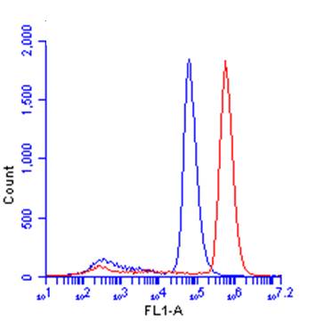
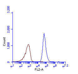
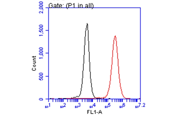
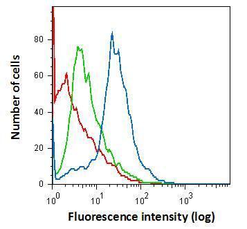
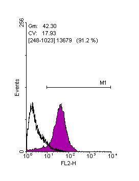
![beta Catenin antibody [N1N2-2], N-term (GRP474) detects CTNNB1 protein by flow cytometry analysis. Sample: HeLa cell. Black: Unlabelled sample was used as a control. Red: beta Catenin antibody [N1N2-2], N-term (GRP474) dilution: 1:50. Acquisition o](https://www.grp-ak.de/media/catalog/product/b/e/beta-catenin-antibody-n1n2-2-n-term_grp474_ihc-p_1_2.jpg)
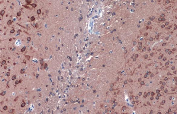
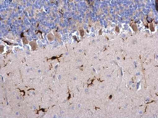

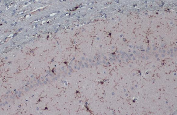

![SOX2 antibody [N1C3] detects SOX2 protein at nucleus on rat brain stem by immunohistochemical analysis. Sample: Paraffin-embedded rat brain stem. SOX2 antibody [N1C3] (GRP557) dilution: 1:500.](https://www.grp-ak.de/media/catalog/product/s/o/sox2-antibody-n1c3_grp557_ihc_6_2.jpg)
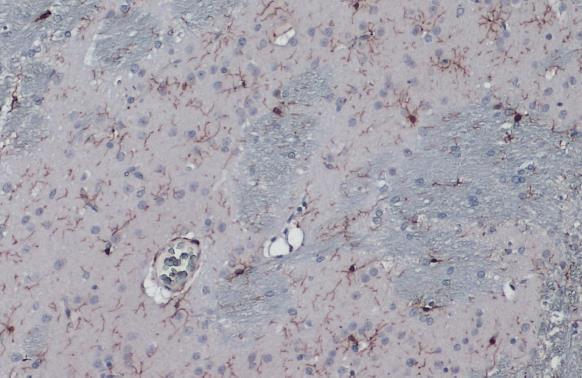
![SOX2 antibody [N1C3] detects SOX2 protein at nucleus on mouse fore brain by immunohistochemical analysis. Sample: Paraffin-embedded mouse fore brain. SOX2 antibody [N1C3] (GRP557) diluted at 1:500.](https://www.grp-ak.de/media/catalog/product/s/o/sox2-antibody-n1c3_grp557_ihc_5_2.jpg)
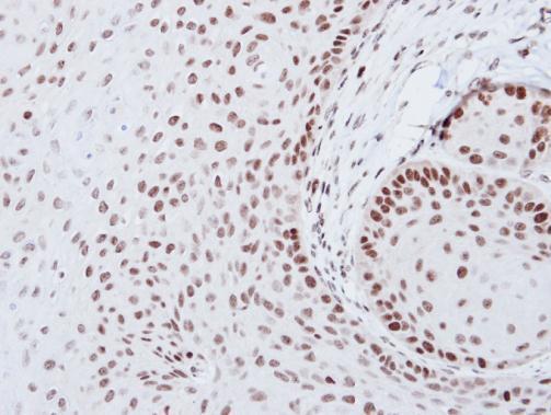
![SOX2 antibody [N1C3] detects SOX2 protein at nucleus in human esophageal carcinoma by immunohistochemical analysis. Sample: Paraffin-embedded human esophageal carcinoma. SOX2 antibody [N1C3] (GRP557) diluted at 1:500.](https://www.grp-ak.de/media/catalog/product/s/o/sox2-antibody-n1c3_grp557_ihc-p_3_2.jpg)
![SOX2 antibody [N1C3] detects SOX2 protein at nucleus in human cervical carcinoma by immunohistochemical analysis. Sample: Paraffin-embedded human cervical carcinoma. SOX2 antibody [N1C3] (GRP557) diluted at 1:500.](https://www.grp-ak.de/media/catalog/product/s/o/sox2-antibody-n1c3_grp557_ihc-p_2_2.jpg)
![SOX2 antibody [N1C3] detects SOX2 protein at nucleus in mouse esophagus by immunohistochemical analysis. Sample: Paraffin-embedded mouse esophagus. SOX2 antibody [N1C3] (GRP557) diluted at 1:500.](https://www.grp-ak.de/media/catalog/product/s/o/sox2-antibody-n1c3_grp557_ihc-p_1_2.jpg)
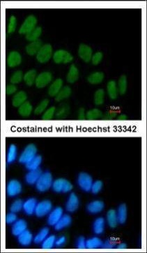
![SOX2 antibody [N1C3] detects SOX2 protein expression by immunohistochemical analysis.Sample: Frozen-sectioned adult mouse hippocampus. Green: SOX2 protein stained by SOX2 antibody [N1C3] (GRP557) diluted at 1:250.Blue: Fluoroshield with DAPI.](https://www.grp-ak.de/media/catalog/product/s/o/sox2-antibody-n1c3_grp557_ihc_4_2.jpg)
![SOX2 antibody [N1C3] detects SOX2 protein at nucleus by immunohistochemical analysis.Sample: Frozen sectioned adult mouse retina. Green: SOX2 protein stained by SOX2 antibody [N1C3] (GRP557) diluted at 1:250.Red: Protein kinase C alpha staining.Blue: Fluo](https://www.grp-ak.de/media/catalog/product/s/o/sox2-antibody-n1c3_grp557_ihc_2_2.jpg)
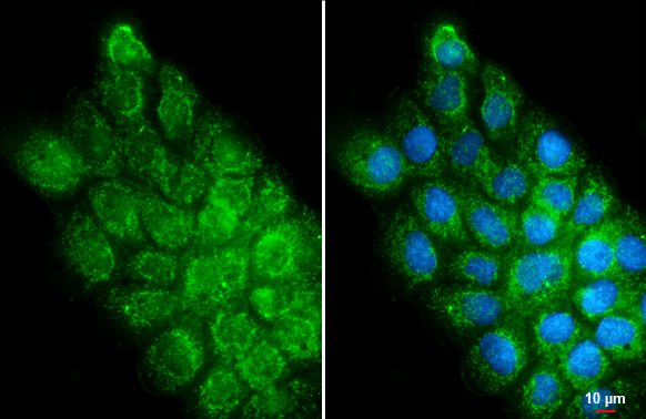
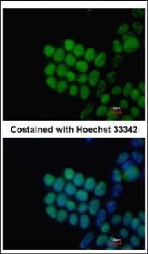
![SOX2 antibody [N1C3] detects SOX2 protein at nucleus by immunofluorescent analysis.Sample: NT2D1 cells were fixed in 4% paraformaldehyde at RT for 15 min.Green: SOX2 stained by SOX2 antibody [N1C3] (GRP557) diluted at 1:500.Red: phalloidin, a cytoskeleton](https://www.grp-ak.de/media/catalog/product/s/o/sox2-antibody-n1c3_grp557_icc_2_2.jpg)
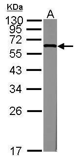
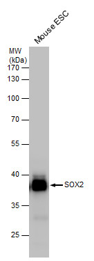
![Whole cell extract (30 μg) was separated by 12% SDS-PAGE, and the membrane was blotted with SOX2 antibody [N1C3] (GRP557) diluted at 1:10000. The HRP-conjugated anti-rabbit IgG antibody was used to detect the primary antibody.](https://www.grp-ak.de/media/catalog/product/s/o/sox2-antibody-n1c3_grp557_wb_2_2.jpg)
![The ICC/IF analysis of SOX2 antibody [N1C3] was published by Chang WF and colleagues in the journal PLoS One in 2016.PMID: 27802323](https://www.grp-ak.de/media/catalog/product/s/o/sox2-antibody-n1c3_grp557_icc_1_2.jpg)
![The WB analysis of SOX2 antibody [N1C3] was published by Misuno K and colleagues in the journal Stem Cell Res Ther in 2013.PMID: 24423398](https://www.grp-ak.de/media/catalog/product/s/o/sox2-antibody-n1c3_grp557_wb_1_2.jpg)
![Immunoprecipitation of SOX2 protein from NT2D1 whole cell extracts using 5 ?g of SOX2 antibody [N1C3] (GRP557).Western blot analysis was performed using SOX2 antibody [N1C3] (GRP557).EasyBlot anti-Rabbit IgG was used as a secondary reagent.](https://www.grp-ak.de/media/catalog/product/s/o/sox2-antibody-n1c3_grp557_ip_2_2.jpg)
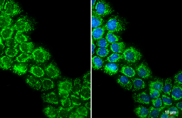
![SOX2 antibody [N1C3] detects SOX2 protein at nucleus in mouse fetal brain by immunohistochemical analysis. Sample: Paraffin-embedded mouse fetal brain. Green: SOX2 antibody [N1C3] (GRP557) diluted at 1:200. The signal was developed using goat anti-rabbit](https://www.grp-ak.de/media/catalog/product/s/o/sox2-antibody-n1c3_grp557_ihc-p_4_2.jpg)
![Immunoprecipitation of SOX2 protein from NT2D1 whole cell extracts using 5 ?g of SOX2 antibody [N1C3] (GRP557) or SOX2 antibody [GT1876] (GRP557).Western blot analysis was performed using SOX2 antibody [N1C3] diluted at 1:500.EasyBlot anti-Rabbit IgG was](https://www.grp-ak.de/media/catalog/product/s/o/sox2-antibody-n1c3_grp557_ip_1_2.jpg)
![SOX2 antibody [N1C3] detects SOX2 protein at nucleus by immunohistochemical analysis.Sample: Frozen sectioned E13.5 rat brain. Green: SOX2 protein stained by SOX2 antibody [N1C3] (GRP557) diluted at 1:250.Red: beta Tubulin 3/ TUJ1, a mature neuron marker,](https://www.grp-ak.de/media/catalog/product/s/o/sox2-antibody-n1c3_grp557_ihc_3_2.jpg)
![Non-transfected (–) and transfected (+) 293T whole cell extracts (30 μg) were separated by 7.5% SDS-PAGE, and the membrane was blotted with GRK2 antibody [C2C3], C-term (GRP559) diluted at 1:1000. The HRP-conjugated anti-rabbit IgG antibody was used](https://www.grp-ak.de/media/catalog/product/g/r/grk2-antibody-c2c3-c-term_grp559_wb_4_2.jpg)
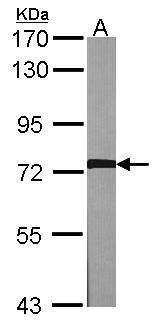
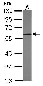
![Various whole cell extracts (30 μg) were separated by 7.5% SDS-PAGE, and the membrane was blotted with GRK2 antibody [C2C3], C-term (GRP559) diluted at 1:1000. The HRP-conjugated anti-rabbit IgG antibody was used to detect the primary antibody.](https://www.grp-ak.de/media/catalog/product/g/r/grk2-antibody-c2c3-c-term_grp559_wb_1_2.jpg)
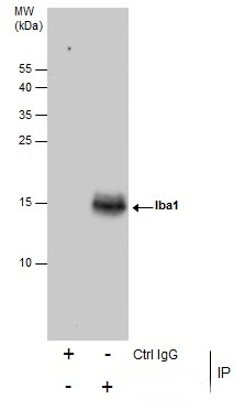
![Immunoprecipitation of GRK2 protein from Jurkat whole cell extracts using 5 ?g of GRK2 antibody [C2C3], C-term (GRP559).Western blot analysis was performed using GRK2 antibody [C2C3], C-term (GRP559).EasyBlot anti-Rabbit IgG was used as a secondary reage](https://www.grp-ak.de/media/catalog/product/g/r/grk2-antibody-c2c3-c-term_grp559_ip_1_2.jpg)