Search results for: 'proteinA'
- 7 images
-
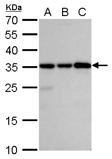
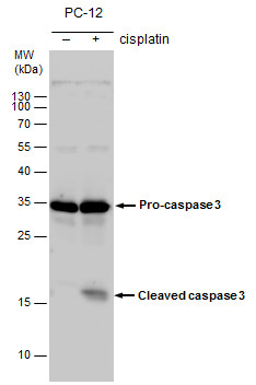
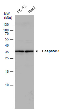
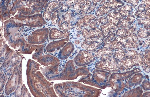
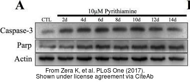
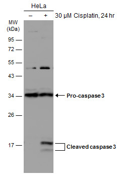
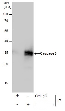
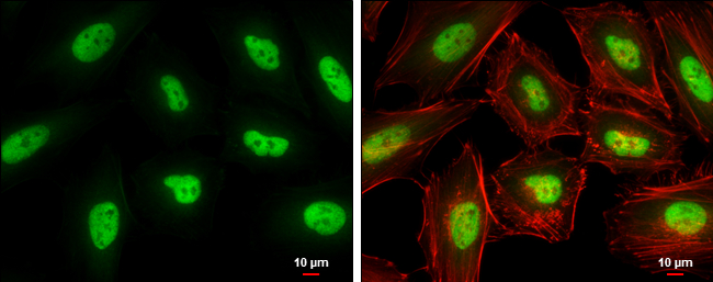
- 10 imagesCaspase 3 antibody [GRP46]
ICC, IF, IHC-Fr, IHC-P, IP, WB
Human, Mouse, Rat
Rabbit
Polyclonal
100 μl -
- 12 imagesRad51 antibody [N1C2] [GRP8]
ICC, IF, IHC-P, IP, WB
Human, Mouse, Rat, Zebrafish
Rabbit
Polyclonal
100 μl -
- Anti-gamma-Tubulin GCP2 Purified [GRP10690]
ICC, IHC-P, IP, WB
Human, Mouse, Rat, Pig
Monoclonal
0.1 mg - 14 images
-
-
- 6 imagesBCL6 antibody [N2C1], Internal [GRP20]
ChIP, IHC-Fr, IHC-P, IP, WB
Human, Mouse, Rat
Rabbit
Polyclonal
100 μl -
-
- 5 imagesMre11 antibody [12D7] [GRP88]
ELISA, FA, ICC, IF, IHC-P, IP, WB
Human, Mouse, Rat
Mouse
Monoclonal
100 μl -
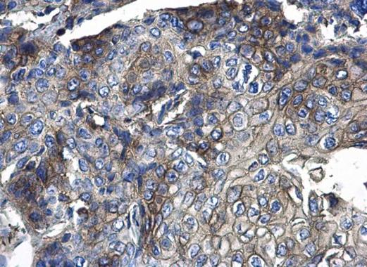
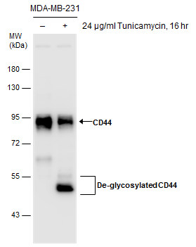
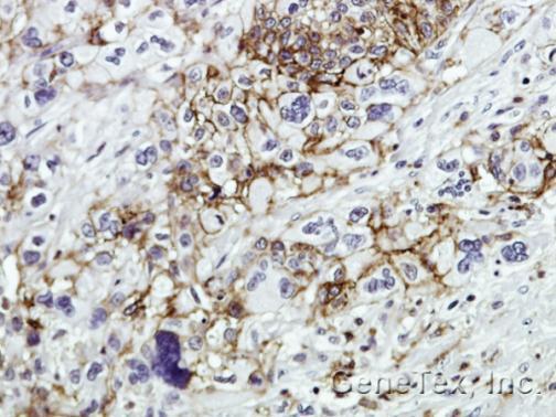
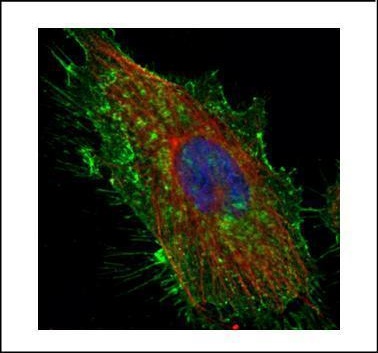
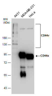
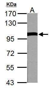
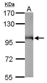
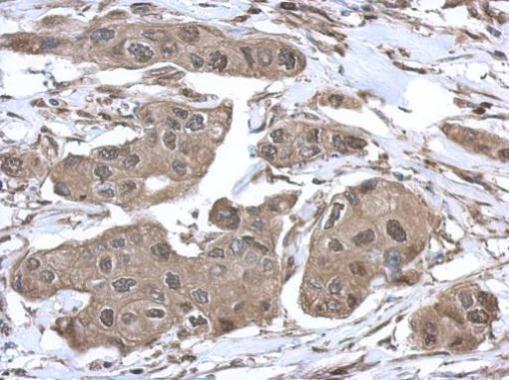
- 5 imagesCD44 antibody [GRP25]
ICC, IF, IHC-Fr, IHC-P, IP, WB
Human, Mouse, Rat, Rabbit
Rabbit
Polyclonal
100 μl -
![VCP antibody detects VCP Protein expression by immunohistochemical analysis.Sample: Frozen-sectioned adult mouse cerebellum. Green: VCP stained by VCP antibody (GRP552) diluted at 1:250.Red: NF-H, stained by NF-H antibody [GT114] (GRP552) diluted at 1:500](https://www.grp-ak.de/media/catalog/product/v/c/vcp-antibody_grp552_ihc_2_2.jpg)
![Various tissue extracts (30 μg) were separated by 10% SDS-PAGE, and the membrane was blotted with Rad51 antibody [N1C2] (GRP460) diluted at 1:500. The HRP-conjugated anti-rabbit IgG antibody was used to detect the primary antibody, and the signal was](https://www.grp-ak.de/media/catalog/product/r/a/rad51-antibody-n1c2_grp460_wb_7_2.jpg)
![Rad51 antibody [N1C2] detects Rad51 protein at nucleus by immunofluorescent analysis.Sample: HeLa cells were fixed in 4% paraformaldehyde at RT for 15 min.Green: Rad51 protein stained by Rad51 antibody [N1C2] (GRP460) diluted at 1:500.Red: phalloidin, a c](https://www.grp-ak.de/media/catalog/product/r/a/rad51-antibody-n1c2_grp460_if_1_2.jpg)
![Various whole cell extracts (30 μg) were separated by 10% SDS-PAGE, and the membrane was blotted with Rad51 antibody [14B4] (GRP460) diluted at 1:500. The HRP-conjugated anti-rabbit IgG antibody was used to detect the primary antibody.](https://www.grp-ak.de/media/catalog/product/r/a/rad51-antibody-n1c2_grp460_wb_6_2.jpg)
![Various whole cell extracts (30 μg) were separated by 10% SDS-PAGE, and the membrane was blotted with Rad51 antibody [N1C2] (GRP460) diluted at 1:1000.](https://www.grp-ak.de/media/catalog/product/r/a/rad51-antibody-n1c2_grp460_wb_5_2.jpg)
![Various whole cell extracts (30 μg) were separated by 10% SDS-PAGE, and the membrane was blotted with Rad51 antibody [14B4] (GRP460) diluted at 1:500. The HRP-conjugated anti-rabbit IgG antibody was used to detect the primary antibody.](https://www.grp-ak.de/media/catalog/product/r/a/rad51-antibody-n1c2_grp460_wb_4_2.jpg)
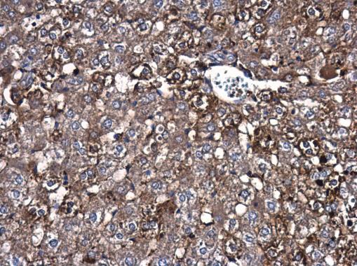
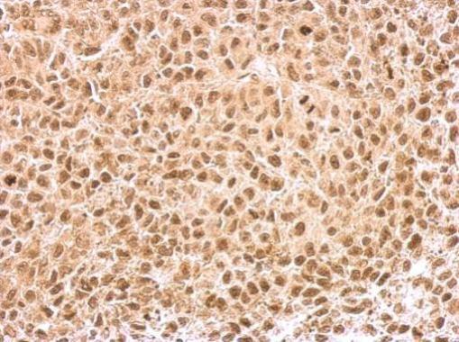
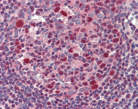
![Rad51 antibody [N1C2] detects Rad51 protein at cytoplasm and nucleus by immunohistochemical analysis.Sample: Paraffin-embedded human cervical carcinoma.Rad51 stained by Rad51 antibody [N1C2] (GRP460) diluted at 1:500.Antigen Retrieval: Citrate buffer, pH](https://www.grp-ak.de/media/catalog/product/r/a/rad51-antibody-n1c2_grp460_ihc-p_3_2.jpg)
![Rad51 antibody [N1C2] detects Rad51 protein at nucleus by immunohistochemical analysis.Sample: Paraffin-embedded mouse testis.Rad51 stained by Rad51 antibody [N1C2] (GRP460) diluted at 1:1000.Antigen Retrieval: Citrate buffer, pH 6.0, 15 min](https://www.grp-ak.de/media/catalog/product/r/a/rad51-antibody-n1c2_grp460_ihc-p_2_2.jpg)
![Rad51 antibody [N1C2] detects Rad51 protein at nucleus by immunohistochemical analysis.Sample: Paraffin-embedded mouse testis.Rad51 stained by Rad51 antibody [N1C2] (GRP460) diluted at 1:500.Antigen Retrieval: Citrate buffer, pH 6.0, 15 min](https://www.grp-ak.de/media/catalog/product/r/a/rad51-antibody-n1c2_grp460_ihc-p_1_2.jpg)
![Untreated (–) and treated (+) HeLa whole cell extracts (30 μg) were separated by 10% SDS-PAGE, and the membrane was blotted with Rad51 antibody [N1C2] (GRP460) diluted at 1:1000. The HRP-conjugated anti-rabbit IgG antibody was used to detect the pri](https://www.grp-ak.de/media/catalog/product/r/a/rad51-antibody-n1c2_grp460_wb_3_2.jpg)
![Various whole cell extracts (30 μg) were separated by 10% SDS-PAGE, and the membrane was blotted with Rad51 antibody [N1C2] (GRP460) diluted at 1:1000. The HRP-conjugated anti-rabbit IgG antibody was used to detect the primary antibody.](https://www.grp-ak.de/media/catalog/product/r/a/rad51-antibody-n1c2_grp460_wb_2_2.jpg)
![Various tissue extracts (30 μg) were separated by 10% SDS-PAGE, and the membrane was blotted with Rad51 antibody [N1C2] (GRP460) diluted at 1:1000. The HRP-conjugated anti-rabbit IgG antibody was used to detect the primary antibody, and the signal was](https://www.grp-ak.de/media/catalog/product/r/a/rad51-antibody-n1c2_grp460_wb_1_2.jpg)
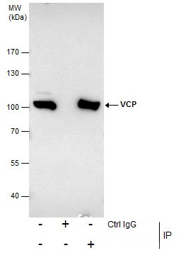
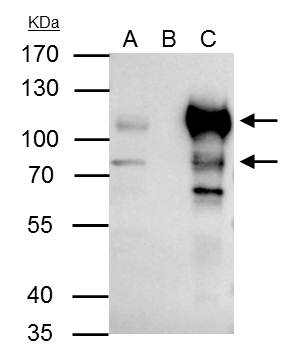
![Rad51 antibody [N1C2] immunoprecipitates Rad51 protein in IP experiments.IP samples: Jurkat whole cell extractA. 40 ?g Jurkat whole cell extractB. Control with 4 ?g of preimmune Rabbit IgGC. Immunoprecipitation of Rad51 protein by 4 ?g Rad51 antibody [N1C](https://www.grp-ak.de/media/catalog/product/r/a/rad51-antibody-n1c2_grp460_ip_1_2.jpg)
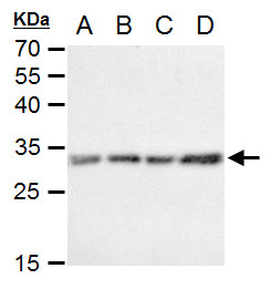
![BCL6 antibody [N2C1], Internal detects BCL6 protein by western blot analysis.A. 30 μg Neuro2A whole cell lysate/extract B. 30 μg GL261 whole cell lysate/extract C. 30 μg C8D30 whole cell lysate/extract D. 30 μg NIH-3T3 whole cell lysate/extrac](https://www.grp-ak.de/media/catalog/product/b/c/bcl6-antibody-n2c1-internal_grp472_wb_2_2.jpg)
![Various whole cell extracts (30 μg) were separated by 7.5% SDS-PAGE, and the membrane was blotted with BCL6 antibody [N2C1], Internal (GRP472) diluted at 1:1000. The HRP-conjugated anti-rabbit IgG antibody was used to detect the primary antibody.](https://www.grp-ak.de/media/catalog/product/b/c/bcl6-antibody-n2c1-internal_grp472_wb_1_2.jpg)
![BCL6 antibody [N2C1], Internal detects BCL6 protein by immunohistochemical analysis.Sample: Frozen-sectioned mouse cerebellum.Green: BCL6 stained by BCL6 antibody [N2C1], Internal (GRP472) diluted at 1:250.Red: NF-H, stained by NF-H antibody [GT114] (GRP4](https://www.grp-ak.de/media/catalog/product/b/c/bcl6-antibody-n2c1-internal_grp472_ihc_2_2.jpg)
![Whole cell extract (30 μg) was separated by 7.5% SDS-PAGE, and the membrane was blotted with Mre11 antibody [12D7] (GRP540) diluted at 1:500. The HRP-conjugated anti-mouse IgG antibody was used to detect the primary antibody, and the signal was develo](https://www.grp-ak.de/media/catalog/product/m/r/mre11-antibody-12d7_grp540_wb_4_2.jpg)
![Mre11 antibody [12D7] detects Mre11 protein by western blot analysis.A. 30 μg 293T whole cell extract B. 30 μg whole cell extract of human Mre11-transfected 293T cells7.5% SDS-PAGEMre11 antibody [12D7] (GRP540) dilution: 1:1000The HRP-conjugated ant](https://www.grp-ak.de/media/catalog/product/m/r/mre11-antibody-12d7_grp540_wb_3_2.jpg)
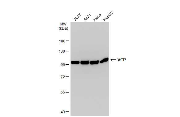
![Various whole cell extracts (30 μg) were separated by 7.5% SDS-PAGE, and the membrane was blotted with Mre11 antibody [12D7] (GRP540) diluted at 1:1000. The HRP-conjugated anti-mouse IgG antibody was used to detect the primary antibody.](https://www.grp-ak.de/media/catalog/product/m/r/mre11-antibody-12d7_grp540_wb_2_2.jpg)
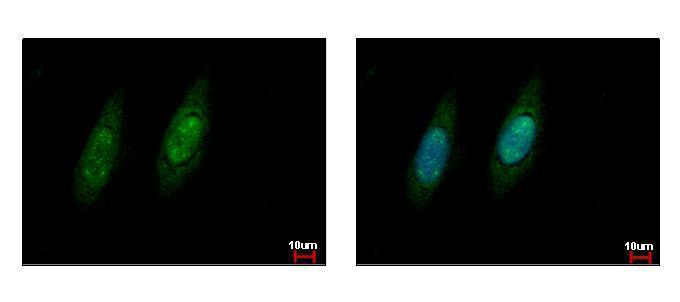
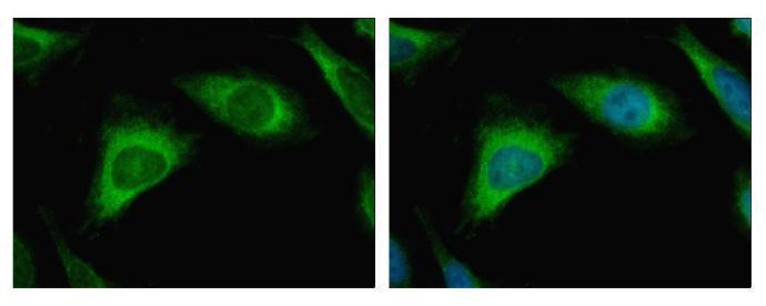
![Mre11 antibody [12D7] detects Mre11 protein at nucleus by immunofluorescent analysis.Sample: HeLa cells were fixed in 4% paraformaldehyde at RT for 15 min.Green: Mre11 stained by Mre11 antibody [12D7] (GRP540) diluted at 1:200.Blue: Hoechst 33342 staining](https://www.grp-ak.de/media/catalog/product/m/r/mre11-antibody-12d7_grp540_icc_1_2.jpg)
![The WB analysis of Mre11 antibody [12D7] was published by Harten SK and colleagues in the journal BMC Biol in 2015.PMID: 25857663](https://www.grp-ak.de/media/catalog/product/m/r/mre11-antibody-12d7_grp540_wb_1_2.jpg)