Search results for: 'proteinA'
-
-
- 2 imagesCathepsin L Polyclonal Antibody [GRP407]
ICC, IF, IHC-P, WB
Human, Mouse, Rat
Rabbit
Polyclonal
100 μl -
- 2 images
-
- 2 images
-
- 6 imagesBCL6 antibody [N2C1], Internal [GRP20]
ChIP, IHC-Fr, IHC-P, IP, WB
Human, Mouse, Rat
Rabbit
Polyclonal
100 μl -
- 2 images
-
- 3 images
-
- 2 images
-
- 3 images
-
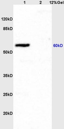
![BCL6 antibody [N2C1], Internal detects BCL6 protein by western blot analysis.A. 30 μg Neuro2A whole cell lysate/extract B. 30 μg GL261 whole cell lysate/extract C. 30 μg C8D30 whole cell lysate/extract D. 30 μg NIH-3T3 whole cell lysate/extrac](https://www.grp-ak.de/media/catalog/product/b/c/bcl6-antibody-n2c1-internal_grp472_wb_2_2.jpg)
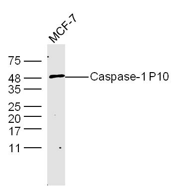
![Various whole cell extracts (30 μg) were separated by 7.5% SDS-PAGE, and the membrane was blotted with BCL6 antibody [N2C1], Internal (GRP472) diluted at 1:1000. The HRP-conjugated anti-rabbit IgG antibody was used to detect the primary antibody.](https://www.grp-ak.de/media/catalog/product/b/c/bcl6-antibody-n2c1-internal_grp472_wb_1_2.jpg)
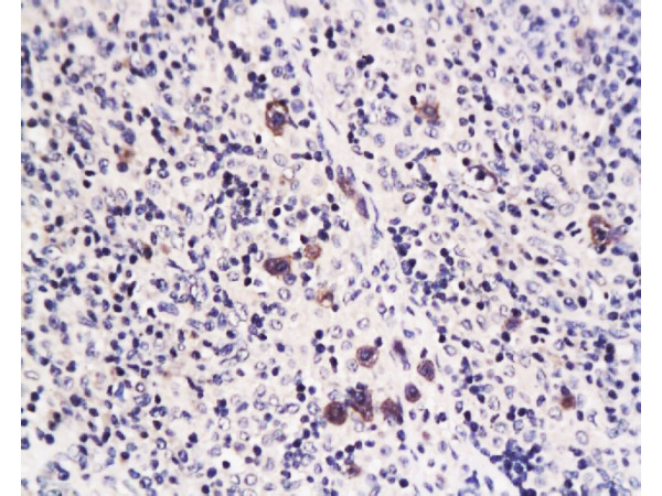
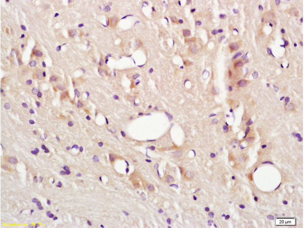
![BCL6 antibody [N2C1], Internal detects BCL6 protein by immunohistochemical analysis.Sample: Frozen-sectioned mouse cerebellum.Green: BCL6 stained by BCL6 antibody [N2C1], Internal (GRP472) diluted at 1:250.Red: NF-H, stained by NF-H antibody [GT114] (GRP4](https://www.grp-ak.de/media/catalog/product/b/c/bcl6-antibody-n2c1-internal_grp472_ihc_2_2.jpg)

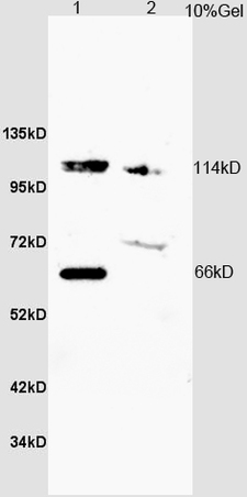
![Whole cell extract (30 μg) was separated by 10% SDS-PAGE, and the membrane was blotted with Opsin 3 antibody [N1], N-term (GRP574) diluted at 1:1000. The HRP-conjugated anti-rabbit IgG antibody was used to detect the primary antibody.](https://www.grp-ak.de/media/catalog/product/o/p/opsin-3-antibody-n1-n-term_grp574_wb_1_2.jpg)
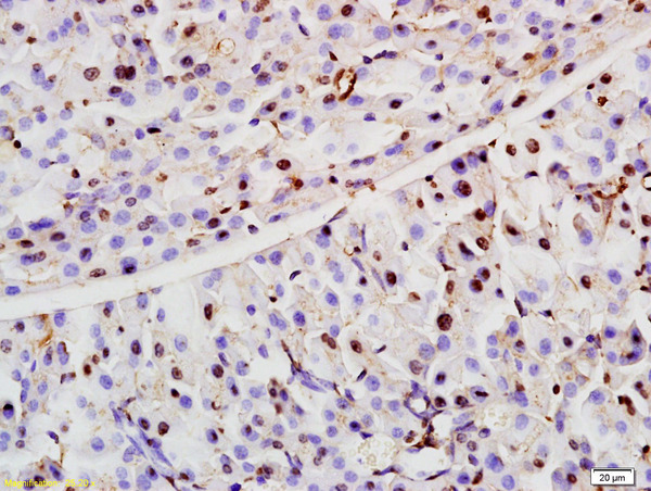
![Opsin 3 antibody [N1], N-term detects Opsin 3 protein expression by immunohistochemical analysis.Sample: Frozen sectioned adult mouse retina. Green: Opsin 3 protein stained by Opsin 3 antibody [N1], N-term (GRP574) diluted at 1:250.Red: beta Tubulin 3/ TU](https://www.grp-ak.de/media/catalog/product/o/p/opsin-3-antibody-n1-n-term_grp574_ihc_2_2.jpg)
