Primary Antibodies
- 15 imagesRad51 antibody [14B4] [GRP90]
ICC, IF, IHC-P, IP, WB
Human, Mouse, Rat, Chicken
Mouse
Monoclonal
100 μl -
- 7 images
-
- 14 images
-
- 3 images
-
- 5 images
-
- 14 images
-
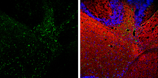
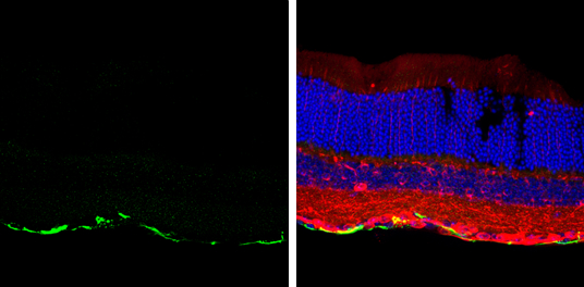
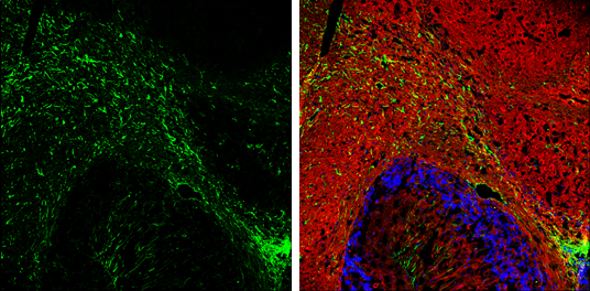
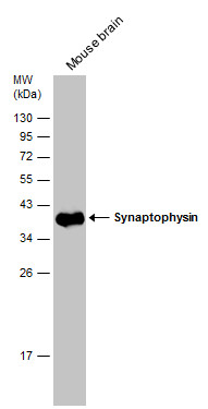
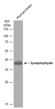
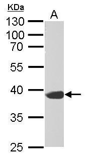
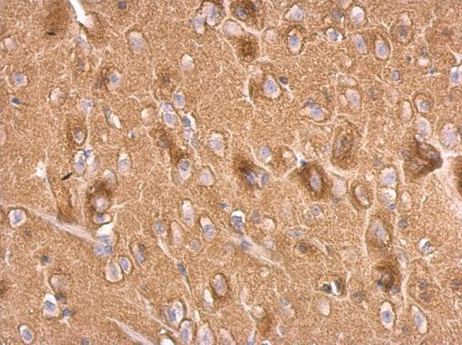
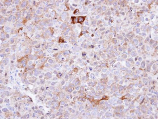
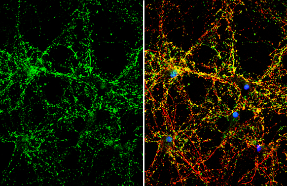
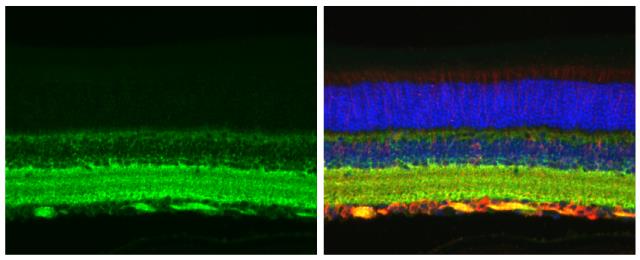
- 10 imagesSynaptophysin antibody [GRP98]
ICC, IF, IHC-Fr, IHC-P, WB
Human, Mouse, Rat
Rabbit
Polyclonal
100 μl -
- 4 images
-
- 7 images
-
- 6 images
-
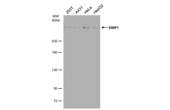
![Various whole cell extracts (30 μg) were separated by 10% SDS-PAGE, and the membrane was blotted with Rad51 antibody [14B4] (GRP542) diluted at 1:500. The HRP-conjugated anti-mouset IgG antibody was used to detect the primary antibody, and the signal](https://www.grp-ak.de/media/catalog/product/r/a/rad51-antibody-14b4_grp542_wb_11_2.jpg)
![Various whole cell extracts (30 μg) were separated by 10% SDS-PAGE, and the membrane was blotted with Rad51 antibody [14B4] (GRP542) diluted at 1:500. The HRP-conjugated anti-mouset IgG antibody was used to detect the primary antibody, and the signal](https://www.grp-ak.de/media/catalog/product/r/a/rad51-antibody-14b4_grp542_wb_10_2.jpg)
![The WB analysis of Rad51 antibody [14B4] was published by Kalimutho M and colleagues in the journal Mol Oncol in 2017 .](https://www.grp-ak.de/media/catalog/product/r/a/rad51-antibody-14b4_grp542_wb_9_2.jpg)
![The WB analysis of Rad51 antibody [14B4] was published by Kalimutho M and colleagues in the journal Mol Oncol in 2017 .](https://www.grp-ak.de/media/catalog/product/r/a/rad51-antibody-14b4_grp542_wb_8_2.jpg)
![The WB analysis of Rad51 antibody [14B4] was published by Kalimutho M and colleagues in the journal Mol Oncol in 2017 .](https://www.grp-ak.de/media/catalog/product/r/a/rad51-antibody-14b4_grp542_wb_7_2.jpg)
![The WB analysis of Rad51 antibody [14B4] was published by Kalimutho M and colleagues in the journal Mol Oncol in 2017 .](https://www.grp-ak.de/media/catalog/product/r/a/rad51-antibody-14b4_grp542_wb_6_2.jpg)
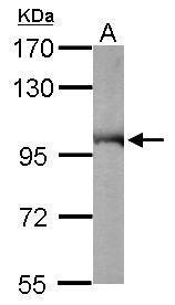
![Various whole cell extracts (30 μg) were separated by 10% SDS-PAGE, and the membrane was blotted with Rad51 antibody [14B4] (GRP542) diluted at 1:500. The HRP-conjugated anti-mouse IgG antibody was used to detect the primary antibody, and the signal w](https://www.grp-ak.de/media/catalog/product/r/a/rad51-antibody-14b4_grp542_wb_5_2.jpg)
![The WB analysis of Rad51 antibody [14B4] was published by Zhu J and colleagues in the journal EMBO Mol Med in 2013.PMID: 23341130](https://www.grp-ak.de/media/catalog/product/r/a/rad51-antibody-14b4_grp542_wb_4_2.jpg)
![The WB analysis of Rad51 antibody [14B4] was published by Zhu J and colleagues in the journal EMBO Mol Med in 2013.PMID: 23341130](https://www.grp-ak.de/media/catalog/product/r/a/rad51-antibody-14b4_grp542_wb_3_2.jpg)
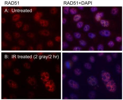
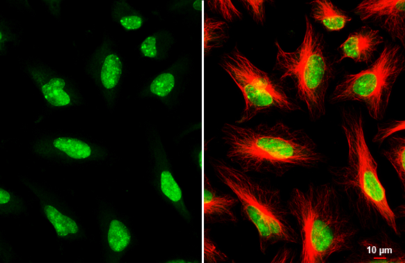
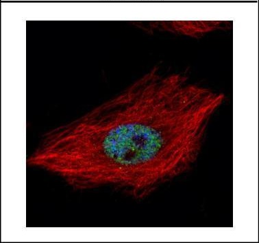
![The ICC/IF analysis of Rad51 antibody [14B4] was published by White MK and colleagues in the journal PLoS One in 2014.PMID: 25310191](https://www.grp-ak.de/media/catalog/product/r/a/rad51-antibody-14b4_grp542_icc_1_2.jpg)
![The WB analysis of Rad51 antibody [14B4] was published by Zhu J and colleagues in the journal EMBO Mol Med in 2013.PMID: 23341130](https://www.grp-ak.de/media/catalog/product/r/a/rad51-antibody-14b4_grp542_wb_2_2.jpg)
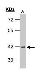
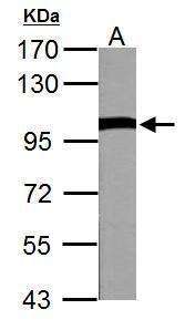
![Whole cell extract (30 μg) was separated by 10% SDS-PAGE, and the membrane was blotted with Rad51 antibody [14B4] (GRP542) diluted at 1:500. The HRP-conjugated anti-mouse IgG antibody was used to detect the primary antibody.](https://www.grp-ak.de/media/catalog/product/r/a/rad51-antibody-14b4_grp542_wb_1_2.jpg)
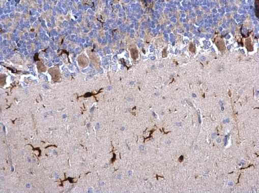
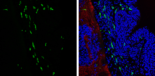

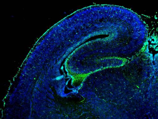
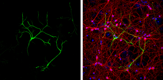
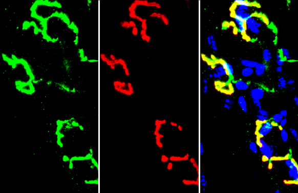
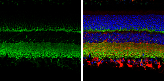
![GFAP antibody detects GFAP protein expression by immunohistochemical analysis.Sample: Frozen-sectioned adult mouse hippocampus. Green: GFAP protein stained by GFAP antibody (GRP549) diluted at 1:250.Red: NeuN, stained by NeuN antibody [2Q158] diluted at](https://www.grp-ak.de/media/catalog/product/g/f/gfap-antibody_grp549_ihc_6_2.jpg)
![GFAP antibodies detects GFAP proteins on embryonic mouse brain by immunohistochemical analysis. Sample: Frozen section of embryonic mouse brain (mE18.5). Green: GFAP antibody (GRP549) diluted at 1:500. Red: Sox2 antibody [GT1876] (GRP549) diluted at 1:500](https://www.grp-ak.de/media/catalog/product/g/f/gfap-antibody_grp549_ihc_3_2.jpg)
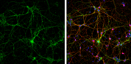
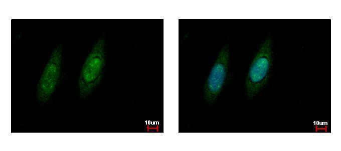
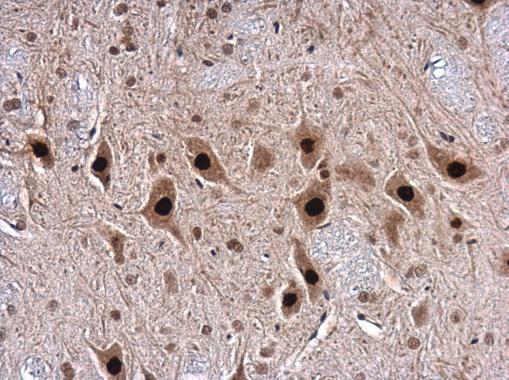
![VCP antibody detects VCP Protein expression by immunohistochemical analysis.Sample: Frozen-sectioned adult mouse cerebellum. Green: VCP stained by VCP antibody (GRP552) diluted at 1:250.Red: NF-H, stained by NF-H antibody [GT114] (GRP552) diluted at 1:500](https://www.grp-ak.de/media/catalog/product/v/c/vcp-antibody_grp552_ihc_2_2.jpg)
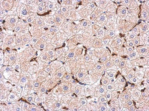
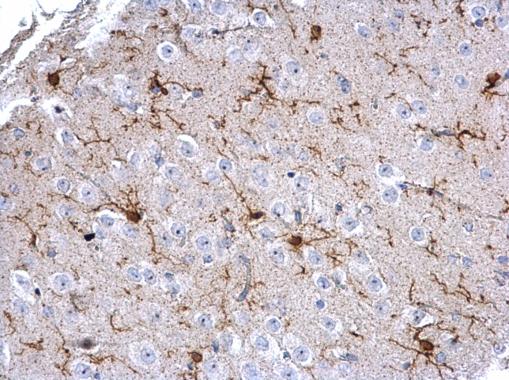
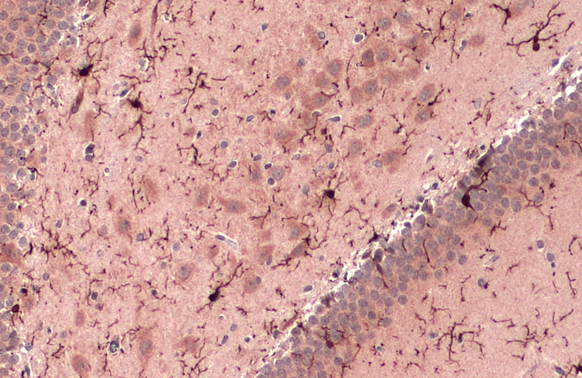
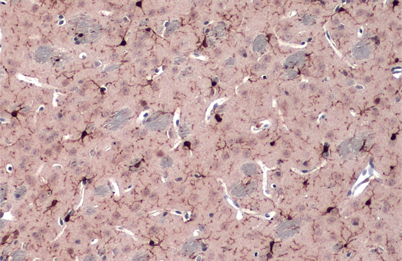
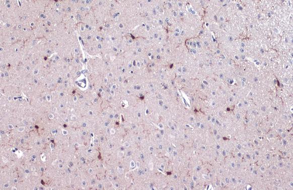
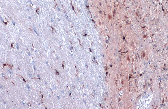
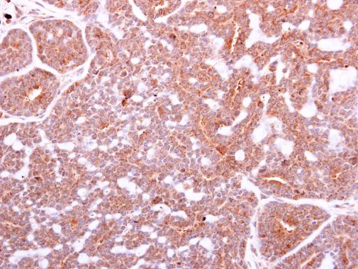
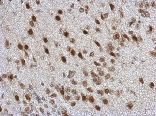
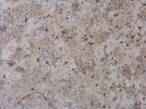
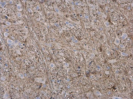
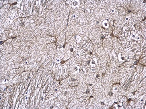
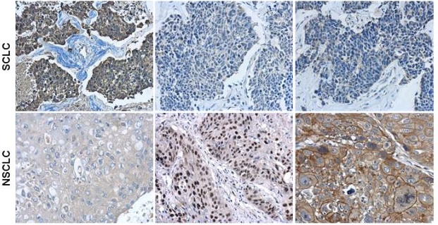
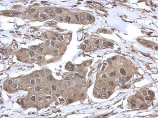
![C3 antibody [C3], C-term detects C3 protein at cytoplasm in mouse brain by immunohistochemical analysis. Sample: Paraffin-embedded mouse brain. C3 antibody [C3], C-term (GRP554) diluted at 1:500.](https://www.grp-ak.de/media/catalog/product/c/3/c3-antibody-c3-c-term_grp554_ihc-p_1_2.jpg)
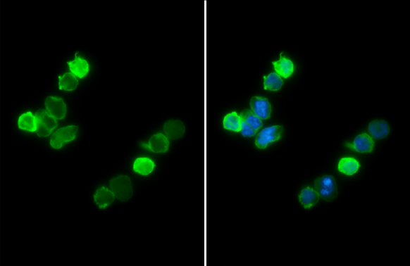
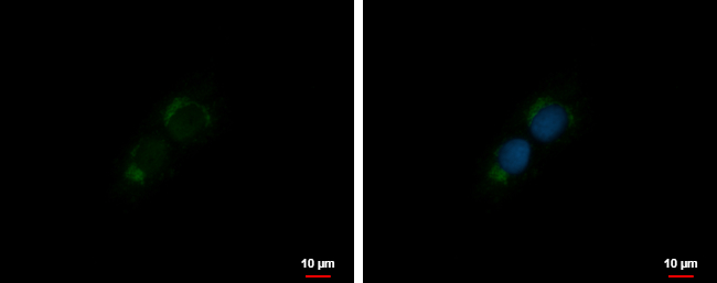
![C3 antibody [C3], C-term detects C3 protein at cytoplasm by immunofluorescent analysis.Sample: HeLa cells were fixed in 4% paraformaldehyde at RT for 15 min.Green: C3 protein stained by C3 antibody [C3], C-term (GRP554) diluted at 1:200.Blue: Hoechst 3334](https://www.grp-ak.de/media/catalog/product/c/3/c3-antibody-c3-c-term_grp554_if_1_2.jpg)

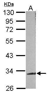
![Human plasma (30 μg) was separated by 7.5% SDS-PAGE, and the membrane was blotted with C3 antibody [C3], C-term (GRP554) diluted at 1:10000. The HRP-conjugated anti-rabbit IgG antibody was used to detect the primary antibody.](https://www.grp-ak.de/media/catalog/product/c/3/c3-antibody-c3-c-term_grp554_wb_2_2.jpg)
![Human plasma (30 μg) was separated by 7.5% SDS-PAGE, and the membrane was blotted with C3 antibody [C3], C-term (GRP554) diluted at 1:10000. The HRP-conjugated anti-rabbit IgG antibody was used to detect the primary antibody.](https://www.grp-ak.de/media/catalog/product/c/3/c3-antibody-c3-c-term_grp554_wb_1_2.jpg)
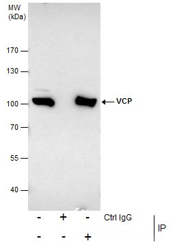
![Immunoprecipitation of C3 protein from HepG2 whole cell extracts using 5 ?g of C3 antibody [C3], C-term (GRP554).Western blot analysis was performed using C3 antibody [C3], C-term (GRP554).EasyBlot anti-Rabbit IgG was used as a secondary reagent.](https://www.grp-ak.de/media/catalog/product/c/3/c3-antibody-c3-c-term_grp554_ip_1_2.jpg)
