Availability
- Request Lead Time
- In stock and ready for quick dispatch
- Usually dispatched within 5-10 working days
Product Overview
| Product Name | Synaptophysin antibody |
|---|---|
| Catalog Number | GRP98 |
| Species/Host | Rabbit |
| Reactivity | Human, Mouse, Rat |
| Conjugation | Unconjugated |
| Tested applications | ICC, IF, IHC-Fr, IHC-P, WB |
| Immunogen | Recombinant protein encompassing a sequence within the C-terminus region of human Synaptophysin. The exact sequence is proprietary. |
| Alternative Names | (click to expand) |
Product Properties
| Form/Appearance | Liquid: 1XPBS, 20% Glycerol (pH7). 0.025% ProClin 300 was added as a preservative. |
|---|---|
| Concentration | 0.25 mg/ml |
| Storage | Store as concentrated solution. Centrifuge briefly prior to opening vial. For short-term storage (1-2 weeks), store at 4°C. For long-term storage, aliquot and store at -20°C or below. Avoid multiple freeze-thaw cycles. |
| Note | For research use only. |
| Isotype | IgG |
| Clonality | Polyclonal |
| Purity | Purified by antigen-affinity chromatography. |
| Uniprot ID | P08247 |
| Entrez | 6855 |
Product Description
Synaptophysin (p38) is an integral membrane protein of small synaptic vesicles in brain and endocrine cells.[supplied by OMIM]
Application Notes
| Dilution Range | WB: 1:500-1:50000,ICC: 1:100-1:1000,IHC-P: 1:100-1:1000 |
|---|
Validation Images
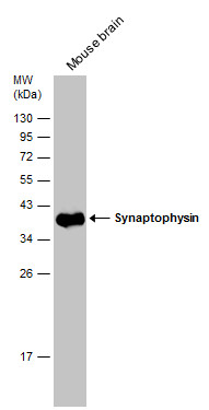
Mouse tissue extract (50 μg) was separated by 12% SDS-PAGE, and the membrane was blotted with Synaptophysin antibody (GRP550) diluted at 1:50000. The HRP-conjugated anti-rabbit IgG antibody was used to detect the primary antibody.
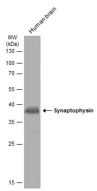
Human tissue extract (30 μg) was separated by 12% SDS-PAGE, and the membrane was blotted with Synaptophysin antibody (GRP550) diluted at 1:500. The HRP-conjugated anti-rabbit IgG antibody was used to detect the primary antibody, and the signal was dev
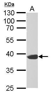
Synaptophysin antibody detects SYP protein by western blot analysis.A. 50 μg rat brain lysate/extract10% SDS-PAGESynaptophysin antibody (GRP550) dilution: 1:10000 The HRP-conjugated anti-rabbit IgG antibody was used to detect the primary antibody.
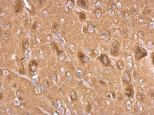
Synaptophysin antibody detects Synaptophysin protein at on rat fore brain by immunohistochemical analysis. Sample: Paraffin-embedded rat fore brain. Synaptophysin antibody (GRP550) dilution: 1:500.
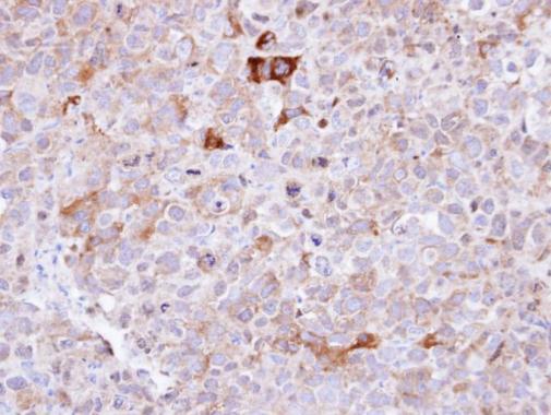
Immunohistochemical analysis of paraffin-embedded CL1-0 xenograft , using Synaptophysin(GRP550) antibody at 1:100 dilution.
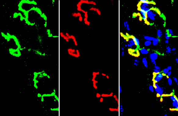
Synaptophysin antibody detects Synaptophysin protein by immunohistochemical analysis.Sample: Frozen-sectioned mouse muscle.Green: Synaptophysin stained by Synaptophysin antibody (GRP550) diluted at ) diluted at 1:5000.Blue: Hoechst 33342 staining.
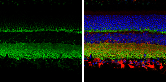
Synaptophysin antibody detects Synaptophysin protein expression by immunohistochemical analysis.Sample: Paraffin-Embedded adult mouse retina. Green: Synaptophysin protein stained by Synaptophysin antibody (GRP550) diluted at 1:250.Red: beta Tubulin 3/ TUJ
Synaptophysin antibody detects Synaptophysin protein at synaptic vesicles by immunofluorescent analysis.Sample: DIV9 rat E18 primary cortical neurons were fixed in 4% paraformaldehyde at RT for 15 min.Green: Synaptophysin protein stained by Synaptophysin
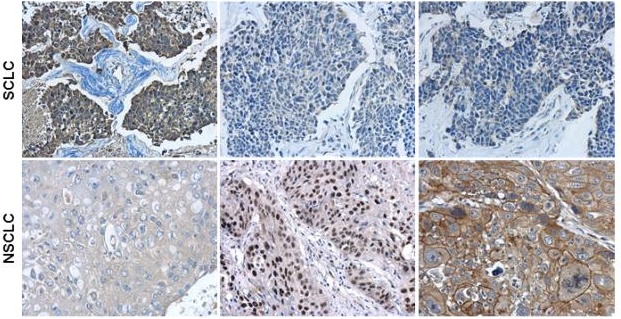
Immunohistochemical characterization of Synaptophysin (GRP550), p63 (GRP550) and Cytokeratin 7 in human small cell lung cancer (SCLC) and non-small cell lung cancer (NSCLC) specimens.Sample: Paraffin-embedded human SCLC (upper panel) and NSCLC (lower pane
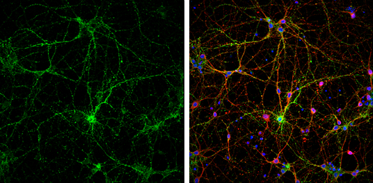
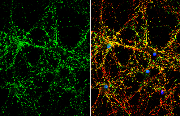
Synaptophysin antibody detects Synaptophysin protein by immunofluorescent analysis.Sample: DIV10 rat E18 primary hippocampal neuron cells were fixed in 4% paraformaldehyde at RT for 15 min.Green: Synaptophysin stained by Synaptophysin antibody (GRP550) di
Reviews
Write Your Own Review
