Primary Antibodies
- 3 images
-
- 7 imagesGRK2 antibody [C2C3], C-term [GRP107]
FACS, ICC, IF, IHC-P, IP, WB
Human, Mouse
Rabbit
Polyclonal
100 μl -
- 10 images
-
- 4 images
-
- 13 imagesTyrosine Hydroxylase antibody [N1C1] [GRP110]
ICC, IF, IHC-Fr, IHC-P, WB
Human, Mouse, Rat, Zebrafish
Rabbit
Polyclonal
100 μl -
- 5 images
-
- 3 images
-
- 8 images
-
- 4 images
-
- 5 images
-
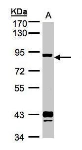
![Non-transfected (–) and transfected (+) 293T whole cell extracts (30 μg) were separated by 7.5% SDS-PAGE, and the membrane was blotted with GRK2 antibody [C2C3], C-term (GRP559) diluted at 1:1000. The HRP-conjugated anti-rabbit IgG antibody was used](https://www.grp-ak.de/media/catalog/product/g/r/grk2-antibody-c2c3-c-term_grp559_wb_4_2.jpg)
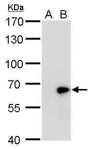
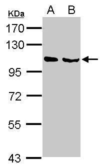
![Various whole cell extracts (30 μg) were separated by 7.5% SDS-PAGE, and the membrane was blotted with GRK2 antibody [C2C3], C-term (GRP559) diluted at 1:1000. The HRP-conjugated anti-rabbit IgG antibody was used to detect the primary antibody.](https://www.grp-ak.de/media/catalog/product/g/r/grk2-antibody-c2c3-c-term_grp559_wb_1_2.jpg)
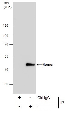
![Immunoprecipitation of GRK2 protein from Jurkat whole cell extracts using 5 ?g of GRK2 antibody [C2C3], C-term (GRP559).Western blot analysis was performed using GRK2 antibody [C2C3], C-term (GRP559).EasyBlot anti-Rabbit IgG was used as a secondary reage](https://www.grp-ak.de/media/catalog/product/g/r/grk2-antibody-c2c3-c-term_grp559_ip_1_2.jpg)

![EMR1 antibody [C2C3], C-term detects EMR1 protein by western blot analysis.A. 50 μg rat liver lysate/extract7.5% SDS-PAGEEMR1 antibody [C2C3], C-term (GRP561) dilution: 1:1000 The HRP-conjugated anti-rabbit IgG antibody was used to detect the primary](https://www.grp-ak.de/media/catalog/product/f/4/f480-antibody-c2c3-c-term_grp561_wb_3_2.jpg)
![Various whole cell extracts (30 μg) were separated by 7.5% SDS-PAGE, and the membrane was blotted with F4/80 antibody [C2C3], C-term (GRP561) diluted at 1:1000. The HRP-conjugated anti-rabbit IgG antibody was used to detect the primary antibody.](https://www.grp-ak.de/media/catalog/product/f/4/f480-antibody-c2c3-c-term_grp561_wb_2_2.jpg)
![Various whole cell extracts (30 μg) were separated by 7.5% SDS-PAGE, and the membrane was blotted with F4/80 antibody [C2C3], C-term (GRP561) diluted at 1:1000. The HRP-conjugated anti-rabbit IgG antibody was used to detect the primary antibody.](https://www.grp-ak.de/media/catalog/product/f/4/f480-antibody-c2c3-c-term_grp561_wb_1_2.jpg)
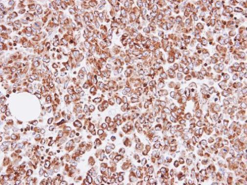
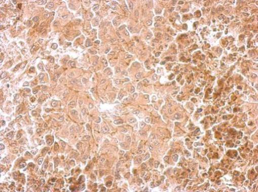
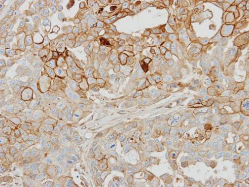
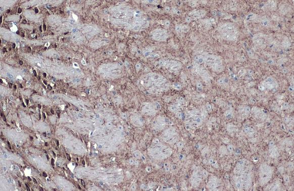
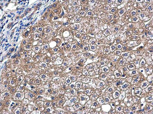
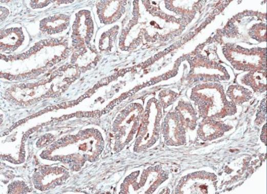
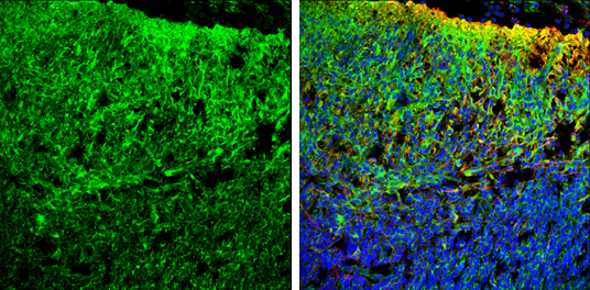
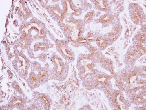
![Tyrosine Hydroxylase antibody [N1C1] detects Tyrosine Hydroxylase protein at cell membrane and cytoplasm by immunohistochemical analysis.Sample: Paraffin-embedded mouse brain.Tyrosine Hydroxylase stained by Tyrosine Hydroxylase antibody [N1C1] (GRP562) di](https://www.grp-ak.de/media/catalog/product/t/y/tyrosine-hydroxylase-antibody-n1c1_grp562_ihc-p_5_2.jpg)
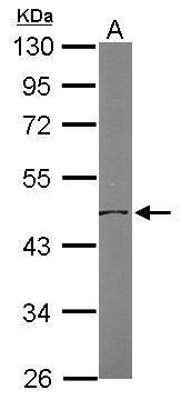
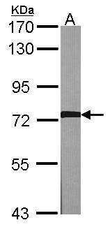
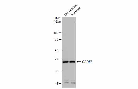
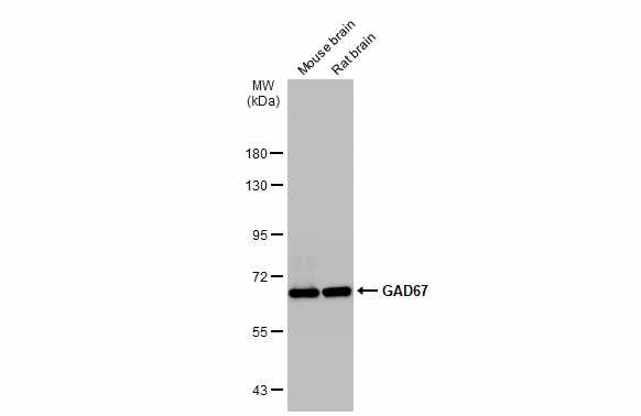
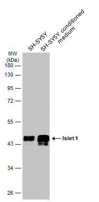
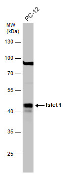
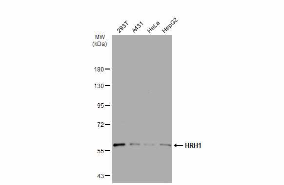
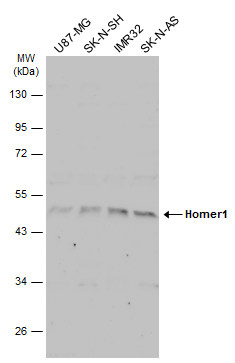
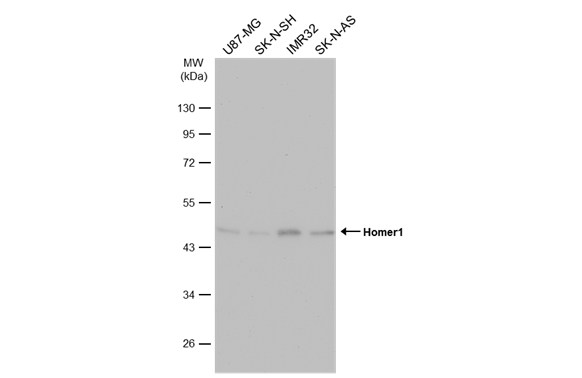
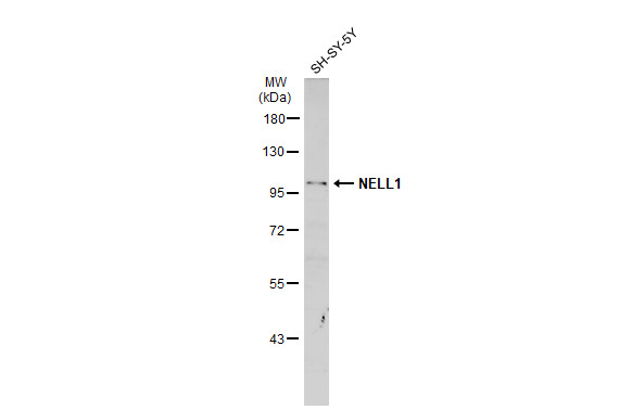
![Whole cell extract (30 μg) was separated by 10% SDS-PAGE, and the membrane was blotted with Tyrosine Hydroxylase antibody [N1C1] (GRP562) diluted at 1:1000. The HRP-conjugated anti-rabbit IgG antibody was used to detect the primary antibody.](https://www.grp-ak.de/media/catalog/product/t/y/tyrosine-hydroxylase-antibody-n1c1_grp562_wb_2_2.jpg)
![Tyrosine Hydroxylase antibody [N1C1] detects Tyrosine Hydroxylase protein on zebrafish by whole mount immunohistochemical analysis. Sample: 2 days-post-fertilization zebrafish embryo. Tyrosine Hydroxylase antibody [N1C1] (GRP562) dilution: 1:100.](https://www.grp-ak.de/media/catalog/product/t/y/tyrosine-hydroxylase-antibody-n1c1_grp562_ihc_3_2.jpg)
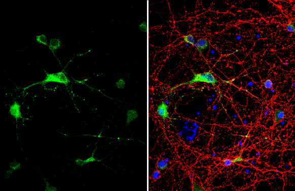
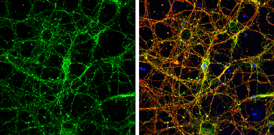
![Tyrosine Hydroxylase antibody [N1C1] detects Tyrosine Hydroxylase protein in midbrain dopaminergic neurons by immunohistochemical analysis.Sample: Paraffin-embedded mouse brain.Green: Tyrosine Hydroxylase stained by Tyrosine Hydroxylase antibody [N1C1] (G](https://www.grp-ak.de/media/catalog/product/t/y/tyrosine-hydroxylase-antibody-n1c1_grp562_ihc-p_4_2.jpg)
![Tyrosine Hydroxylase antibody [N1C1] detects Tyrosine Hydroxylase protein on zebrafish by whole mount immunohistochemical analysis. Sample: 2 days-post-fertilization zebrafish embryo. Tyrosine Hydroxylase antibody [N1C1] (GRP562) dilution: 1:100.](https://www.grp-ak.de/media/catalog/product/t/y/tyrosine-hydroxylase-antibody-n1c1_grp562_ihc_1_2.jpg)
![Tyrosine Hydroxylase antibody [N1C1] detects Tyrosine Hydroxylase protein at cell membrane and cytoplasm by immunohistochemical analysis.Sample: Paraffin-embedded mouse brain.Tyrosine Hydroxylase stained by Tyrosine Hydroxylase antibody [N1C1] (GRP562) di](https://www.grp-ak.de/media/catalog/product/t/y/tyrosine-hydroxylase-antibody-n1c1_grp562_ihc-p_3_2.jpg)
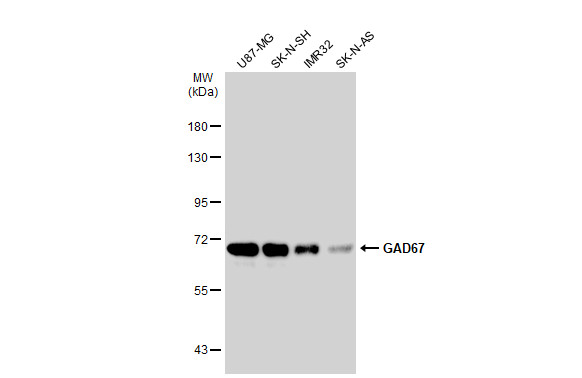
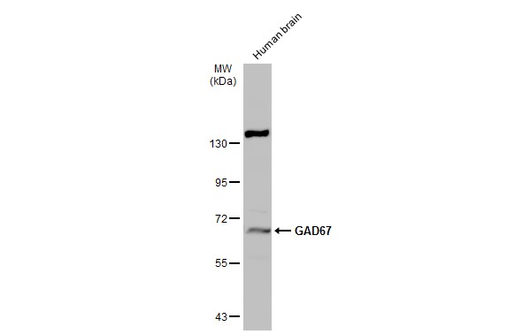
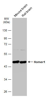
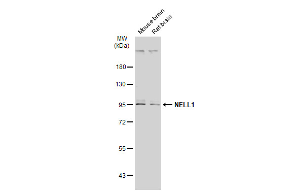
![Various tissue extracts (50 μg) were separated by 10% SDS-PAGE, and the membrane was blotted with Tyrosine Hydroxylase antibody [N1C1] (GRP562) diluted at 1:1000. The HRP-conjugated anti-rabbit IgG antibody was used to detect the primary antibody.](https://www.grp-ak.de/media/catalog/product/t/y/tyrosine-hydroxylase-antibody-n1c1_grp562_wb_1_2.jpg)
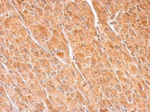
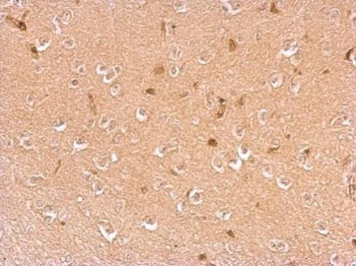
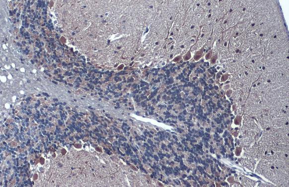
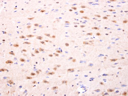
![Tyrosine Hydroxylase antibody [N1C1] detects Tyrosine Hydroxylase protein at cytoplasm by immunohistochemical analysis.Sample: Paraffin-embedded mouse brain.Tyrosine Hydroxylase stained by Tyrosine Hydroxylase antibody [N1C1] (GRP562) diluted at 1:1000.An](https://www.grp-ak.de/media/catalog/product/t/y/tyrosine-hydroxylase-antibody-n1c1_grp562_ihc-p_2_2.jpg)
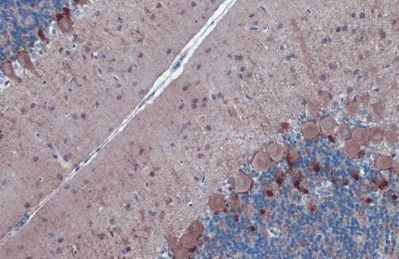
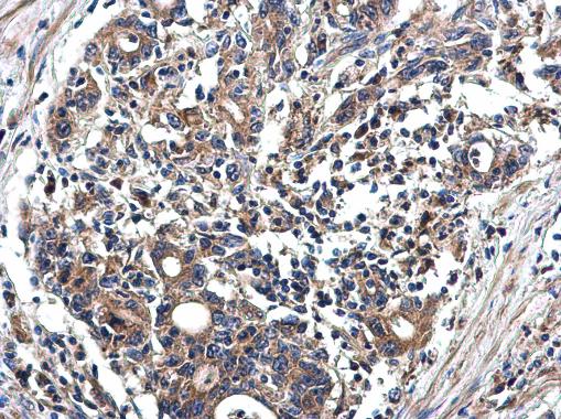
![Tyrosine Hydroxylase antibody [N1C1] detects Tyrosine Hydroxylase protein at cytoplasm by immunohistochemical analysis.Sample: Paraffin-embedded rat brain.Tyrosine Hydroxylase stained by Tyrosine Hydroxylase antibody [N1C1] (GRP562) diluted at 1:1000.Anti](https://www.grp-ak.de/media/catalog/product/t/y/tyrosine-hydroxylase-antibody-n1c1_grp562_ihc-p_1_2.jpg)
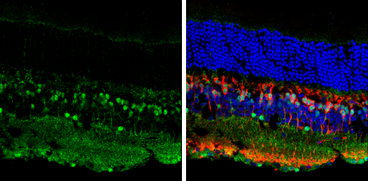
![Tyrosine Hydroxylase antibody [N1C1] detects Tyrosine Hydroxylase protein by immunohistochemical analysis.Sample: Frozen sectioned adult mouse retina. Green: Tyrosine Hydroxylase protein stained by Tyrosine Hydroxylase antibody [N1C1] (GRP562) diluted at](https://www.grp-ak.de/media/catalog/product/t/y/tyrosine-hydroxylase-antibody-n1c1_grp562_ihc_4_2.jpg)
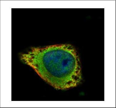
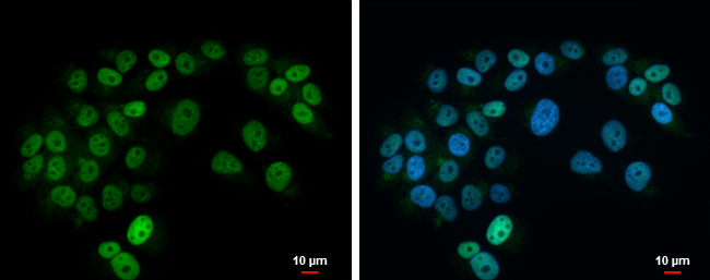
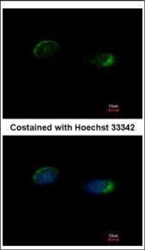
![Tyrosine Hydroxylase antibody [N1C1] detects Tyrosine Hydroxylase protein at cytoplasm by immunofluorescent analysis.Sample: HeLa cells were fixed in 4% paraformaldehyde at RT for 15 min.Green: Tyrosine Hydroxylase protein stained by Tyrosine Hydroxylase](https://www.grp-ak.de/media/catalog/product/t/y/tyrosine-hydroxylase-antibody-n1c1_grp562_if_2_2.jpg)
![Tyrosine Hydroxylase antibody [N1C1] detects Tyrosine Hydroxylase protein expression by immunohistochemical analysis.Sample: Frozen sectioned E13.5 Rat brain. Green: Tyrosine Hydroxylase protein stained by Tyrosine Hydroxylase antibody [N1C1] (GRP562) dil](https://www.grp-ak.de/media/catalog/product/t/y/tyrosine-hydroxylase-antibody-n1c1_grp562_ihc_2_2.jpg)
![Tyrosine Hydroxylase antibody [N1C1] detects Tyrosine Hydroxylase protein at cytoplasm by immunofluorescent analysis.Sample: SK-N-SH cells were fixed in 4% paraformaldehyde at RT for 15 min.Green: Tyrosine Hydroxylase protein stained by Tyrosine Hydroxyla](https://www.grp-ak.de/media/catalog/product/t/y/tyrosine-hydroxylase-antibody-n1c1_grp562_if_1_2.jpg)
![Various whole cell extracts (30 μg) were separated by 10% SDS-PAGE, and the membrane was blotted with OAT antibody [N1C3] (GRP567) diluted at 1:1000. The HRP-conjugated anti-rabbit IgG antibody was used to detect the primary antibody.](https://www.grp-ak.de/media/catalog/product/o/a/oat-antibody-n1c3_grp567_wb_3_2.jpg)
![OAT antibody [N1C3] detects OAT protein at mitochondria by immunofluorescent analysis.Sample: HeLa cells were fixed in ice-cold MeOH for 5 min.Green: OAT protein stained by OAT antibody [N1C3] (GRP567) diluted at 1:500.Blue: Hoechst 33342 staining.](https://www.grp-ak.de/media/catalog/product/o/a/oat-antibody-n1c3_grp567_if_1_2.jpg)
![Non-transfected (–) and transfected (+) 293T whole cell extracts (30 μg) were separated by 10% SDS-PAGE, and the membrane was blotted with OAT antibody [N1C3] (GRP567) diluted at 1:5000. The HRP-conjugated anti-rabbit IgG antibody was used to detect](https://www.grp-ak.de/media/catalog/product/o/a/oat-antibody-n1c3_grp567_wb_2_2.jpg)