Search results for: 'Hepatitis B virus X Protein'
- 1 imageHepatitis Virus (Strain A59) Nonstructural Protein 9 (nsp9) (MOUSE) Monoclonal Antibody [GRP2086]
IF, WB
Mouse
Mouse
Monoclonal
100ug -
- 9 imagesHistone H2A.XS139ph (phospho Ser139) antibody [GT2311] [GRP79]
ICC, IF, IHC-P, IP, WB
Human, Mouse, Rat
Mouse
Monoclonal
100 μl -
-
-
-
- 2 images
-
- Anti-RPSA Purified [GRP10429]
ELISA, ICC, IHC-P, IP, WB
Human, Mouse, Bovine, Chicken, Frog
Monoclonal
0.1 mg - 8 images
-
- 4 images
-
- 12 imagesRad50 antibody [13B3] [GRP89]
ICC, IF, IHC-P, IP, WB
Human, Mouse, Rat, Monkey
Mouse
Monoclonal
100 μl -
![Histone H2A.XS139ph (phospho Ser139) antibody [GT2311] detects Histone H2A.XS139ph (phospho Ser139) protein at nucleus on mouse testis by immunohistochemical analysis. Sample: Paraffin-embedded mouse testis. Histone H2A.XS139ph (phospho Ser139) antibody [](https://www.grp-ak.de/media/catalog/product/h/i/histone-h2a.xs139ph-phospho-ser139-antibody-gt2311_grp531_ihc_1_2.jpg)
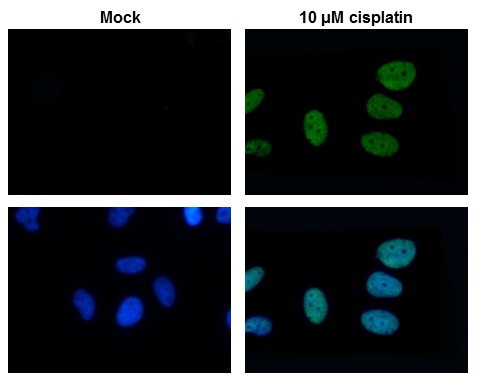
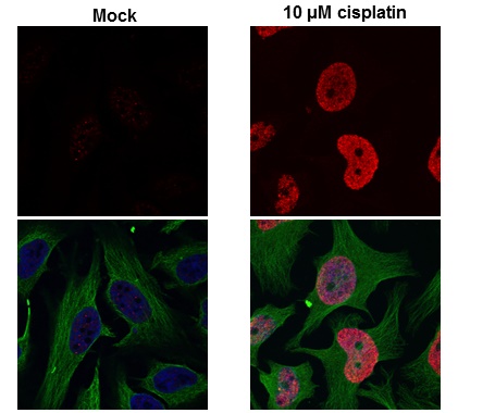
![Histone H2A.X (phospho S139) antibody [GT2311] detects H2AFX protein by western blot analysis.A. 30 μg NIH-3T3 whole cell lysate/extract (untreated)B. 30 μg NIH-3T3 whole cell lysate/extract (30μM cisplatin treatment for 24hr)15% SDS-PAGEHistone](https://www.grp-ak.de/media/catalog/product/h/i/histone-h2a.xs139ph-phospho-ser139-antibody-gt2311_grp531_wb_5_2.jpg)
![Histone H2A.X (phospho S139) antibody [GT2311] detects Histone H2A.X (phospho S139) [GT2311] protein by western blot analysis. Un-treated (-) and treated (+, 30 μM Cisplatin treatment for 24 hrs) PC-12 whole cell extracts (30 μg) were separated by 1](https://www.grp-ak.de/media/catalog/product/h/i/histone-h2a.xs139ph-phospho-ser139-antibody-gt2311_grp531_wb_4_2.jpg)
![Histone H2A.X (phospho S139) antibody [GT2311] detects H2AFX protein by western blot analysis.A. 30 μg HCT116 whole cell lysate/extract (untreated)B. 30 μg HCT116 whole cell lysate/extract (30 μM cisplatin treatment for 24hr)12% SDS-PAGEHistone H](https://www.grp-ak.de/media/catalog/product/h/i/histone-h2a.xs139ph-phospho-ser139-antibody-gt2311_grp531_wb_2_2.jpg)
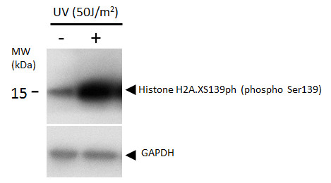
![The WB analysis of Histone H2A.XS139ph (phospho Ser139) antibody [GT2311] was published by En J and colleagues in the journal PLoS Negl Trop Dis in 2017.PMID: 28783752](https://www.grp-ak.de/media/catalog/product/h/i/histone-h2a.xs139ph-phospho-ser139-antibody-gt2311_grp531_wb_1_2.jpg)
![TDP43 antibody [GT225] detects TDP43 protein at nucleus in rat brain by immunohistochemical analysis. Sample: Paraffin-embedded rat brain. TDP43 antibody [GT225] (GRP624) diluted at 1:200.](https://www.grp-ak.de/media/catalog/product/t/d/tdp43-antibody-gt225_grp624_ihc-p_2_2.jpg)
![TARDBP antibody [GT225] detects TARDBP protein by western blot analysis.A. 30 μg 293T whole cell lysate/extract B. 30 μg A431 whole cell lysate/extract C. 30 μg HeLa whole cell lysate/extract10 % SDS-PAGETARDBP antibody [GT225] (GRP624) dilution:](https://www.grp-ak.de/media/catalog/product/t/d/tdp43-antibody-gt225_grp624_wb_4_2.jpg)
![TDP43 antibody [GT225] detects TDP43 protein at nucleus in mouse brain by immunohistochemical analysis. Sample: Paraffin-embedded mouse brain. TDP43 antibody [GT225] (GRP624) diluted at 1:200.](https://www.grp-ak.de/media/catalog/product/t/d/tdp43-antibody-gt225_grp624_ihc-p_1_2.jpg)
![TARDBP antibody [GT225] detects TARDBP protein by western blot analysis.A. 30 μg BCL-1 whole cell lysate/extract B. 30 μg Raw264.7 whole cell lysate/extract10 % SDS-PAGETARDBP antibody [GT225] (GRP624) dilution: 1:1000](https://www.grp-ak.de/media/catalog/product/t/d/tdp43-antibody-gt225_grp624_wb_3_2.jpg)
![TARDBP antibody [GT225] detects TARDBP protein by western blot analysis.A. 30 μg PC-12 whole cell lysate/extractB. 30 μg Rat2 whole cell lysate/extract10 % SDS-PAGETARDBP antibody [GT225] (GRP624) dilution: 1:1000](https://www.grp-ak.de/media/catalog/product/t/d/tdp43-antibody-gt225_grp624_wb_2_2.jpg)
![Various whole cell extracts (30 μg) were separated by 10% SDS-PAGE, and the membrane was blotted with TARDBP antibody [GT225] (GRP624) diluted at 1:500.](https://www.grp-ak.de/media/catalog/product/t/d/tdp43-antibody-gt225_grp624_wb_1_2.jpg)
![TDP43 antibody [GT225] detects TDP43 protein by immunofluorescent analysis.Sample: DIV10 rat E18 primary cortical neuron cells were fixed in 4% paraformaldehyde at RT for 15 min.Green: Nestin stained by Nestin antibody (GRP624) diluted at 1:500.Red: TDP43](https://www.grp-ak.de/media/catalog/product/t/d/tdp43-antibody-gt225_grp624_icc_2_2.jpg)
![TDP43 antibody [GT225] detects TDP43 protein immunofluorescent analysis.Sample: DIV10 rat E18 primary cortical neuron cells were fixed in 4% paraformaldehyde at RT for 15 min.Green: Nestin stained by Nestin antibody (GRP624) diluted at 1:500.Red: TDP43 st](https://www.grp-ak.de/media/catalog/product/t/d/tdp43-antibody-gt225_grp624_icc_1_2.jpg)
![Rat tissue extract (50 μg) was separated by 10% SDS-PAGE, and the membrane was blotted with LAMP1 antibody [GT25212] (GRP628) diluted at 1:1000. The HRP-conjugated anti-mouse IgG antibody was used to detect the primary antibody, and the signal was dev](https://www.grp-ak.de/media/catalog/product/l/a/lamp1-antibody-gt25212_grp628_wb_1_2.jpg)
![LAMP1 antibody [GT25212] detects LAMP1 protein at cytoplasm by immunohistochemical analysis.Sample: Paraffin-embedded mouse liver.LAMP1 stained by LAMP1 antibody [GT25212] (GRP628) diluted at 1:1000.Antigen Retrieval: Citrate buffer, pH 6.0, 15 min](https://www.grp-ak.de/media/catalog/product/l/a/lamp1-antibody-gt25212_grp628_ihc-p_2_2.jpg)
![LAMP1 antibody [GT25212] detects LAMP1 protein at cytoplasm by immunohistochemical analysis.Sample: Paraffin-embedded rat liver.LAMP1 stained by LAMP1 antibody [GT25212] (GRP628) diluted at 1:1000.Antigen Retrieval: Citrate buffer, pH 6.0, 15 min](https://www.grp-ak.de/media/catalog/product/l/a/lamp1-antibody-gt25212_grp628_ihc-p_1_2.jpg)
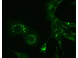
![LAMP1 antibody [GT25212] detects LAMP1 protein at lysosome by immunofluorescent analysis.Sample: HeLa cells were fixed in ice-cold MeOH for 5 min.Green: LAMP1 stained by LAMP1 antibody [GT25212] (GRP628) diluted at 1:2000.Red: alpha Tubulin 4a, a cytoskel](https://www.grp-ak.de/media/catalog/product/l/a/lamp1-antibody-gt25212_grp628_icc_1_2.jpg)
![Rad50 antibody [13B3] detects Rad50 protein at nucleus by immunofluorescent analysis.Sample: HeLa cells were fixed in 4% paraformaldehyde at RT for 15 min.Green: Rad50 protein stained by Rad50 antibody [13B3] (GRP541) diluted at 1:200.Red: phalloidin, a c](https://www.grp-ak.de/media/catalog/product/r/a/rad50-antibody-13b3_grp541_if_1_2.jpg)
![HeLa whole cell and nuclear extracts (30 μg) were separated by 5% SDS-PAGE, and the membrane was blotted with Rad50 antibody [13B3] (GRP541) diluted at 1:1000. The HRP-conjugated anti-mouset IgG antibody was used to detect the primary antibody.](https://www.grp-ak.de/media/catalog/product/r/a/rad50-antibody-13b3_grp541_wb_6_2.jpg)
![Rad50 antibody [13B3] detects Rad50 protein at nucleus in CAL 27 xenograft by immunohistochemical analysis. Sample: Paraffin-embedded CAL 27 xenograft. Rad50 antibody [13B3] (GRP541) diluted at 1:200.](https://www.grp-ak.de/media/catalog/product/r/a/rad50-antibody-13b3_grp541_ihc-p_5_2.jpg)
![Rad50 antibody [13B3] detects Rad50 protein at nucleus in human lung by immunohistochemical analysis. Sample: Paraffin-embedded human lung. Rad50 antibody [13B3] (GRP541) diluted at 1:200.](https://www.grp-ak.de/media/catalog/product/r/a/rad50-antibody-13b3_grp541_ihc-p_4_2.jpg)
![Rad50 antibody [13B3] detects Rad50 protein at nucleus in PC-3 xenograft by immunohistochemical analysis. Sample: Paraffin-embedded PC-3 xenograft. Rad50 antibody [13B3] (GRP541) diluted at 1:200.](https://www.grp-ak.de/media/catalog/product/r/a/rad50-antibody-13b3_grp541_ihc-p_3_2.jpg)
![Rad50 antibody [13B3] detects Rad50 protein at nucleus by immunohistochemical analysis.Sample: Paraffin-embedded human lung cancer.Rad50 stained by Rad50 antibody [13B3] (GRP541) diluted at 1:100.Antigen Retrieval: Citrate buffer, pH 6.0, 15 min](https://www.grp-ak.de/media/catalog/product/r/a/rad50-antibody-13b3_grp541_ihc-p_2_2.jpg)
![Rad50 antibody [13B3] detects Rad50 protein at nucleus by immunohistochemical analysis.Sample: Paraffin-embedded human lung cancer.Rad50 stained by Rad50 antibody [13B3] (GRP541) diluted at 1:100.Antigen Retrieval: Citrate buffer, pH 6.0, 15 min](https://www.grp-ak.de/media/catalog/product/r/a/rad50-antibody-13b3_grp541_ihc-p_1_2.jpg)
![The WB analysis of Rad50 antibody [13B3] was published by Palagyi A and colleagues in the journal Mol Cancer in 2010 .](https://www.grp-ak.de/media/catalog/product/r/a/rad50-antibody-13b3_grp541_wb_5_2.jpg)
![The WB, IP analysis of Rad50 antibody [13B3] was published by Mariggiò G and colleagues in the journal PLoS Pathog in 2017.PMID: 28430817](https://www.grp-ak.de/media/catalog/product/r/a/rad50-antibody-13b3_grp541_wb_4_2.jpg)
![The WB analysis of Rad50 antibody [13B3] was published by Mariggiò G and colleagues in the journal PLoS Pathog in 2017.PMID: 28430817](https://www.grp-ak.de/media/catalog/product/r/a/rad50-antibody-13b3_grp541_wb_3_2.jpg)
![The WB analysis of Rad50 antibody [13B3] was published by Zhu J and colleagues in the journal EMBO Mol Med in 2013.PMID: 23341130](https://www.grp-ak.de/media/catalog/product/r/a/rad50-antibody-13b3_grp541_wb_2_2.jpg)
![The WB analysis of Rad50 antibody [13B3] was published by Harten SK and colleagues in the journal BMC Biol in 2015.PMID: 25857663](https://www.grp-ak.de/media/catalog/product/r/a/rad50-antibody-13b3_grp541_wb_1_2.jpg)
