Search results for: 'Formyl peptide ant'
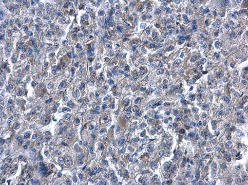
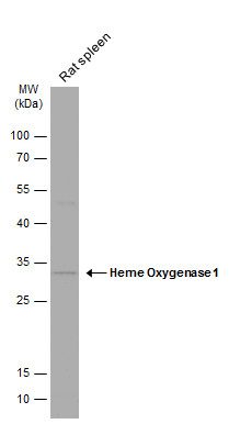
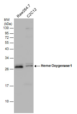
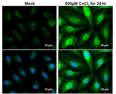
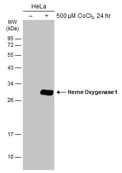
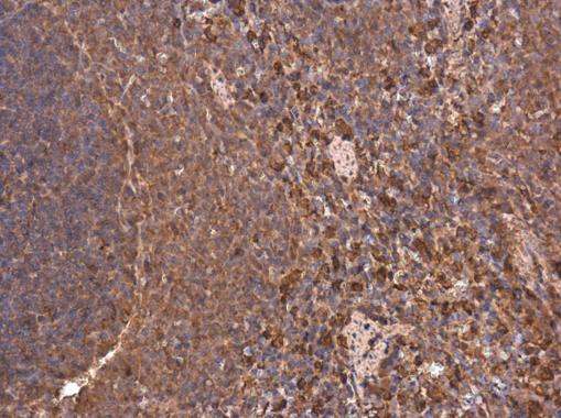
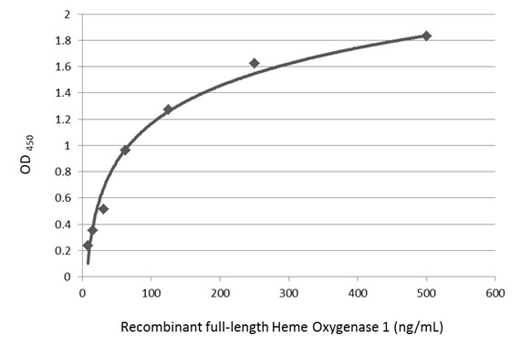
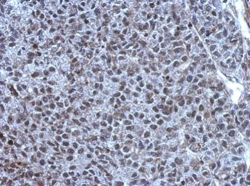
- 7 imagesHeme Oxygenase 1 antibody [GRP19]
ELISA, ICC, IF, IHC-P, WB
Human, Mouse, Rat, Monkey
Rabbit
Polyclonal
100 μl -
- 7 imagesTET1 antibody [N3C1] [GRP63]
ChIP, ICC, IF, IHC-P, IP, WB
Human, Mouse, Monkey
Rabbit
Polyclonal
100 μl -
- 10 imagesATM antibody [2C1] [GRP83]
ChIP, ELISA, FACS, ICC, IF, IHC-P, IP, WB
Human, Mouse, Rat, Monkey
Mouse
Monoclonal
100 μl -
- 10 imagesEstrogen Receptor beta antibody [14C8] [GRP87]
ChIP, DOT, FACS, ICC, IF, IHC-P, WB
Human, Mouse, Monkey
Mouse
Monoclonal
100 μl -
- 12 imagesRad50 antibody [13B3] [GRP89]
ICC, IF, IHC-P, IP, WB
Human, Mouse, Rat, Monkey
Mouse
Monoclonal
100 μl -
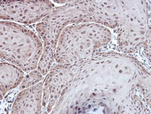
- 7 imagesAspartoacylase antibody [N1C3-2] [GRP138]
ICC, IF, IHC-P, WB
Human, Mouse, Monkey
Rabbit
Polyclonal
100 μl -
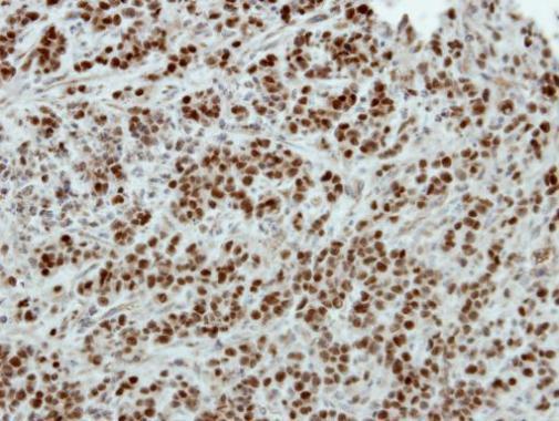
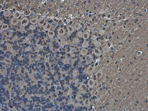
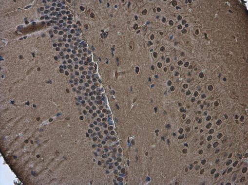
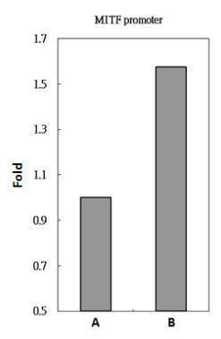
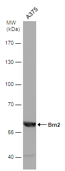
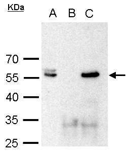
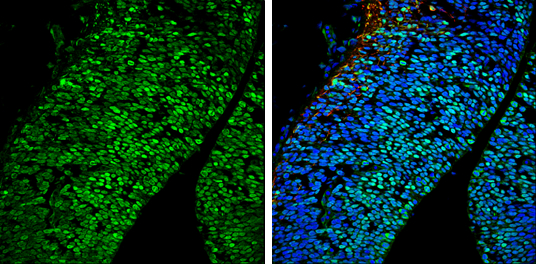
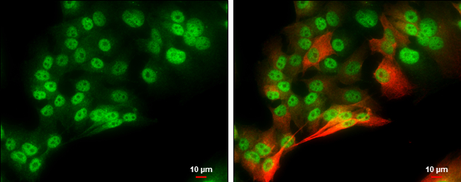
- 8 imagesBrn2 antibody [GRP144]
ChIP, ICC, IF, IHC-Fr, IHC-P, IP, WB
Human, Mouse, Rat, Monkey
Rabbit
Polyclonal
100 μl -
-
![Various whole cell extracts (30 μg) were separated by 5% SDS-PAGE, and the membrane was blotted with TET1 antibody [N3C1] (GRP515) diluted at 1:2000. The HRP-conjugated anti-rabbit IgG antibody was used to detect the primary antibody.](https://www.grp-ak.de/media/catalog/product/t/e/tet1-antibody-n3c1_grp515_wb_3_2.jpg)
![TET1 antibody [N3C1] detects TET1 protein at nucleus in human A549 xenograft by immunohistochemical analysis. Sample: Paraffin-embedded human A549 xenograft . TET1 antibody [N3C1] (GRP515) diluted at 1:250.](https://www.grp-ak.de/media/catalog/product/t/e/tet1-antibody-n3c1_grp515_ihc-p_1_2.jpg)
![TET1 antibody [N3C1] detects TET1 protein at nucleus on Human normal prostate tissue by immunohistochemical analysis. Sample: Paraffin-embedded Human normal prostate tissue. TET1 antibody [N3C1] (GRP515) dilution: 1:1000.](https://www.grp-ak.de/media/catalog/product/t/e/tet1-antibody-n3c1_grp515_ihc_2_2.jpg)
![HeLa whole cell and nuclear extracts (30 μg) were separated by 5% SDS-PAGE, and the membrane was blotted with TET1 antibody [N3C1] (GRP515) diluted at 1:1000. The HRP-conjugated anti-rabbit IgG antibody was used to detect the primary antibody.](https://www.grp-ak.de/media/catalog/product/t/e/tet1-antibody-n3c1_grp515_wb_2_2.jpg)
![TET1 antibody [N3C1] detects TET1 protein at nucleus by immunofluorescent analysis.Sample: Mock and transfected 293T cells were fixed in 4% paraformaldehyde at RT for 15 min.Green: TET1 stained by TET1 antibody [N3C1] (GRP515) diluted at 1:1000.Blue: Hoec](https://www.grp-ak.de/media/catalog/product/t/e/tet1-antibody-n3c1_grp515_icc_1_2.jpg)
![TET1 antibody [N3C1] detects TET1 protein by western blot analysis.A. 30 μg 293T whole cell lysate/extractB. 30 μg whole cell lysate/extract of DDDDK-human TET1-transfected 293T cells5% SDS-PAGETET1 antibody [N3C1] (GRP515) dilution: 1:5000 The HRP-](https://www.grp-ak.de/media/catalog/product/t/e/tet1-antibody-n3c1_grp515_wb_1_2.jpg)
![Whole cell extract (30 μg) was separated by 5% SDS-PAGE, and the membrane was blotted with ATM antibody [2C1] (GRP535) diluted at 1:1000.](https://www.grp-ak.de/media/catalog/product/a/t/atm-antibody-2c1_grp535_wb_6_2.jpg)
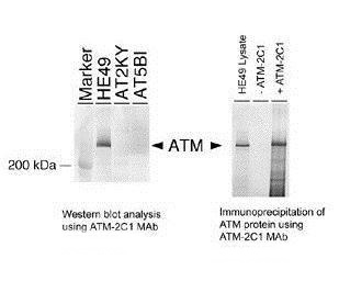
![HeLa whole cell extract and nuclear extracts (30 μg) were separated by 5% SDS-PAGE, and the membrane was blotted with ATM antibody [2C1] (GRP535) diluted at 1:500. The HRP-conjugated anti-mouse IgG antibody was used to detect the primary antibody.](https://www.grp-ak.de/media/catalog/product/a/t/atm-antibody-2c1_grp535_wb_5_2.jpg)
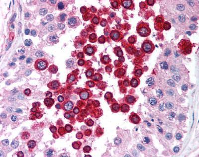
![ATM antibody [2C1] detects ATM protein at nucleus by immunohistochemical analysis.Sample: Paraffin-embedded human breast carcinoma.ATM stained by ATM antibody [2C1] (GRP535) diluted at 1:100.Antigen Retrieval: Citrate buffer, pH 6.0, 15 min](https://www.grp-ak.de/media/catalog/product/a/t/atm-antibody-2c1_grp535_ihc-p_1_2.jpg)
![The WB analysis of ATM antibody [2C1] was published by Lee JH and colleagues in the journal PLoS One in 2014 .](https://www.grp-ak.de/media/catalog/product/a/t/atm-antibody-2c1_grp535_wb_4_2.jpg)
![The WB analysis of ATM antibody [2C1] was published by Kongruttanachok N and colleagues in the journal Mol Cancer in 2010.PMID: 20356374](https://www.grp-ak.de/media/catalog/product/a/t/atm-antibody-2c1_grp535_wb_3_2.jpg)
![The WB analysis of ATM antibody [2C1] was published by He D and colleagues in the journal Sci Rep in 2016.PMID: 27074761](https://www.grp-ak.de/media/catalog/product/a/t/atm-antibody-2c1_grp535_wb_2_2.jpg)
![The WB analysis of ATM antibody [2C1] was published by Gibbs-Seymour I and colleagues in the journal Aging Cell in 2015.PMID: 25645366](https://www.grp-ak.de/media/catalog/product/a/t/atm-antibody-2c1_grp535_wb_1_2.jpg)
![Non-transfected (–) and transfected (+) 293T whole cell extracts (30 μg) were separated by 7.5% SDS-PAGE, and the membrane was blotted with Estrogen Receptor beta antibody [14C8] (GRP539) diluted at 1:5000. The HRP-conjugated anti-mouse IgG antibody](https://www.grp-ak.de/media/catalog/product/e/s/estrogen-receptor-beta-antibody-14c8_grp539_wb_6_2.jpg)
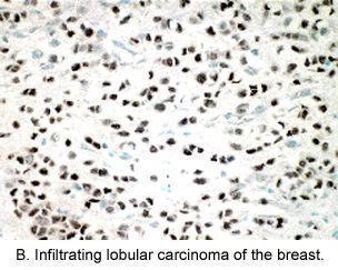
![Estrogen Receptor beta antibody [14C8] detects Estrogen Receptor beta protein at nucleus by immunohistochemical analysis.Sample: Paraffin-embedded human breast carcinoma.Estrogen Receptor beta stained by Estrogen Receptor beta antibody [14C8] (GRP539) dil](https://www.grp-ak.de/media/catalog/product/e/s/estrogen-receptor-beta-antibody-14c8_grp539_ihc-p_3_2.jpg)
![The WB analysis of Estrogen Receptor beta antibody [14C8] was published by Thomas C and colleagues in the journal Breast Cancer Res in 2012 .](https://www.grp-ak.de/media/catalog/product/e/s/estrogen-receptor-beta-antibody-14c8_grp539_wb_5_2.jpg)
![The WB analysis of Estrogen Receptor beta antibody [14C8] was published by Thomas C and colleagues in the journal Breast Cancer Res in 2012 .](https://www.grp-ak.de/media/catalog/product/e/s/estrogen-receptor-beta-antibody-14c8_grp539_wb_4_2.jpg)
![The WB analysis of Estrogen Receptor beta antibody [14C8] was published by Thomas C and colleagues in the journal Breast Cancer Res in 2012 .](https://www.grp-ak.de/media/catalog/product/e/s/estrogen-receptor-beta-antibody-14c8_grp539_wb_3_2.jpg)
![The WB analysis of Estrogen Receptor beta antibody [14C8] was published by Thomas C and colleagues in the journal Breast Cancer Res in 2012 .](https://www.grp-ak.de/media/catalog/product/e/s/estrogen-receptor-beta-antibody-14c8_grp539_wb_2_2.jpg)
![The WB analysis of Estrogen Receptor beta antibody [14C8] was published by Thomas C and colleagues in the journal Breast Cancer Res in 2012 .](https://www.grp-ak.de/media/catalog/product/e/s/estrogen-receptor-beta-antibody-14c8_grp539_wb_1_2.jpg)
![The IHC-P analysis of Estrogen Receptor beta antibody [14C8] was published by Samartzis N and colleagues in the journal Reprod Biol Endocrinol in 2012.PMID: 22520060](https://www.grp-ak.de/media/catalog/product/e/s/estrogen-receptor-beta-antibody-14c8_grp539_ihc-p_2_2.jpg)
![The IHC-P analysis of Estrogen Receptor beta antibody [14C8] was published by Hata S and colleagues in the journal Cancer Med in 2013.PMID: 23930207](https://www.grp-ak.de/media/catalog/product/e/s/estrogen-receptor-beta-antibody-14c8_grp539_ihc-p_1_2.jpg)
![Rad50 antibody [13B3] detects Rad50 protein at nucleus by immunofluorescent analysis.Sample: HeLa cells were fixed in 4% paraformaldehyde at RT for 15 min.Green: Rad50 protein stained by Rad50 antibody [13B3] (GRP541) diluted at 1:200.Red: phalloidin, a c](https://www.grp-ak.de/media/catalog/product/r/a/rad50-antibody-13b3_grp541_if_1_2.jpg)
![HeLa whole cell and nuclear extracts (30 μg) were separated by 5% SDS-PAGE, and the membrane was blotted with Rad50 antibody [13B3] (GRP541) diluted at 1:1000. The HRP-conjugated anti-mouset IgG antibody was used to detect the primary antibody.](https://www.grp-ak.de/media/catalog/product/r/a/rad50-antibody-13b3_grp541_wb_6_2.jpg)
![Rad50 antibody [13B3] detects Rad50 protein at nucleus in CAL 27 xenograft by immunohistochemical analysis. Sample: Paraffin-embedded CAL 27 xenograft. Rad50 antibody [13B3] (GRP541) diluted at 1:200.](https://www.grp-ak.de/media/catalog/product/r/a/rad50-antibody-13b3_grp541_ihc-p_5_2.jpg)
![Rad50 antibody [13B3] detects Rad50 protein at nucleus in human lung by immunohistochemical analysis. Sample: Paraffin-embedded human lung. Rad50 antibody [13B3] (GRP541) diluted at 1:200.](https://www.grp-ak.de/media/catalog/product/r/a/rad50-antibody-13b3_grp541_ihc-p_4_2.jpg)
![Rad50 antibody [13B3] detects Rad50 protein at nucleus in PC-3 xenograft by immunohistochemical analysis. Sample: Paraffin-embedded PC-3 xenograft. Rad50 antibody [13B3] (GRP541) diluted at 1:200.](https://www.grp-ak.de/media/catalog/product/r/a/rad50-antibody-13b3_grp541_ihc-p_3_2.jpg)
![Rad50 antibody [13B3] detects Rad50 protein at nucleus by immunohistochemical analysis.Sample: Paraffin-embedded human lung cancer.Rad50 stained by Rad50 antibody [13B3] (GRP541) diluted at 1:100.Antigen Retrieval: Citrate buffer, pH 6.0, 15 min](https://www.grp-ak.de/media/catalog/product/r/a/rad50-antibody-13b3_grp541_ihc-p_2_2.jpg)
![Rad50 antibody [13B3] detects Rad50 protein at nucleus by immunohistochemical analysis.Sample: Paraffin-embedded human lung cancer.Rad50 stained by Rad50 antibody [13B3] (GRP541) diluted at 1:100.Antigen Retrieval: Citrate buffer, pH 6.0, 15 min](https://www.grp-ak.de/media/catalog/product/r/a/rad50-antibody-13b3_grp541_ihc-p_1_2.jpg)
![The WB analysis of Rad50 antibody [13B3] was published by Palagyi A and colleagues in the journal Mol Cancer in 2010 .](https://www.grp-ak.de/media/catalog/product/r/a/rad50-antibody-13b3_grp541_wb_5_2.jpg)
![The WB, IP analysis of Rad50 antibody [13B3] was published by Mariggiò G and colleagues in the journal PLoS Pathog in 2017.PMID: 28430817](https://www.grp-ak.de/media/catalog/product/r/a/rad50-antibody-13b3_grp541_wb_4_2.jpg)
![The WB analysis of Rad50 antibody [13B3] was published by Mariggiò G and colleagues in the journal PLoS Pathog in 2017.PMID: 28430817](https://www.grp-ak.de/media/catalog/product/r/a/rad50-antibody-13b3_grp541_wb_3_2.jpg)
![The WB analysis of Rad50 antibody [13B3] was published by Zhu J and colleagues in the journal EMBO Mol Med in 2013.PMID: 23341130](https://www.grp-ak.de/media/catalog/product/r/a/rad50-antibody-13b3_grp541_wb_2_2.jpg)
![The WB analysis of Rad50 antibody [13B3] was published by Harten SK and colleagues in the journal BMC Biol in 2015.PMID: 25857663](https://www.grp-ak.de/media/catalog/product/r/a/rad50-antibody-13b3_grp541_wb_1_2.jpg)
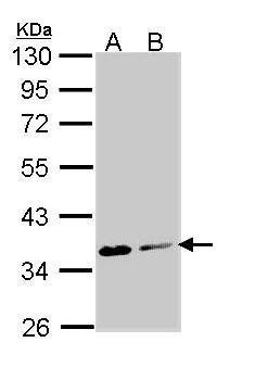
![Non-transfected (–) and transfected (+) 293T whole cell extracts (30 μg) were separated by 10% SDS-PAGE, and the membrane was blotted with Aspartoacylase antibody [N1C3-2] (GRP590) diluted at 1:10000. The HRP-conjugated anti-rabbit IgG antibody was](https://www.grp-ak.de/media/catalog/product/a/s/aspartoacylase-antibody-n1c3-2_grp590_wb_4_2.jpg)
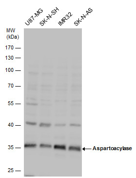
![Various whole cell extracts (30 μg) were separated by 10% SDS-PAGE, and the membrane was blotted with Aspartoacylase antibody [N1C3-2] (GRP590) diluted at 1:1000. The HRP-conjugated anti-rabbit IgG antibody was used to detect the primary antibody.](https://www.grp-ak.de/media/catalog/product/a/s/aspartoacylase-antibody-n1c3-2_grp590_wb_2_2.jpg)
![Various tissue extracts (50 μg) were separated by 10% SDS-PAGE, and the membrane was blotted with Aspartoacylase antibody [N1C3-2] (GRP590) diluted at 1:1000. The HRP-conjugated anti-rabbit IgG antibody was used to detect the primary antibody.](https://www.grp-ak.de/media/catalog/product/a/s/aspartoacylase-antibody-n1c3-2_grp590_wb_1_2.jpg)
![Aspartoacylase antibody [N1C3-2] detects Aspartoacylase protein at cytoplasm by immunofluorescent analysis.Sample: HeLa cells were fixed in 4% paraformaldehyde at RT for 15 min.Green: Aspartoacylase protein stained by Aspartoacylase antibody [N1C3-2] (GRP](https://www.grp-ak.de/media/catalog/product/a/s/aspartoacylase-antibody-n1c3-2_grp590_if_1_2.jpg)