Availability
- Request Lead Time
- In stock and ready for quick dispatch
- Usually dispatched within 5-10 working days
Product Overview
| Product Name | N-Cadherin antibody |
|---|---|
| Catalog Number | GRP68 |
| Species/Host | Rabbit |
| Reactivity | Human, Mouse, Rat |
| Conjugation | Unconjugated |
| Tested applications | ICC, IF, IHC-Fr, IHC-P, WB |
| Immunogen | Carrier-protein conjugated synthetic peptide encompassing a sequence within the C-terminus region of human N-Cadherin. The exact sequence is proprietary. |
| Alternative Names | (click to expand) |
Product Properties
| Form/Appearance | Liquid: 1XPBS, 1% BSA, 20% Glycerol (pH7). 0.025% ProClin 300 was added as a preservative. |
|---|---|
| Concentration | 0.48 mg/ml |
| Storage | Store as concentrated solution. Centrifuge briefly prior to opening vial. For short-term storage (1-2 weeks), store at 4°C. For long-term storage, aliquot and store at -20°C or below. Avoid multiple freeze-thaw cycles. |
| Note | For research use only. |
| Isotype | IgG |
| Clonality | Polyclonal |
| Purity | Purified by antigen-affinity chromatography. |
| Uniprot ID | P19022 |
| Entrez | 1000 |
Product Description
This gene is a classical cadherin from the cadherin superfamily. The encoded protein is a calcium dependent cell-cell adhesion glycoprotein comprised of five extracellular cadherin repeats, a transmembrane region and a highly conserved cytoplasmic tail. The protein functions during gastrulation and is required for establishment of left-right asymmetry. At certain central nervous system synapses, presynaptic to postsynaptic adhesion is mediated at least in part by this gene product. [provided by RefSeq]
Application Notes
| Dilution Range | WB: 1:500-1:3000,ICC: 1:100-1:1000,IHC-P: 1:100-1:1000,IHC-Fr: 1:100-1:1000 |
|---|
Validation Images
N-Cadherin antibody detects N-Cadherin protein on embryonic mouse brain by immunohistochemical analysis. Sample: Frozen section of embryonic mouse brain (mE18.5). N-Cadherin antibody (GRP520) diluted at 1:500.
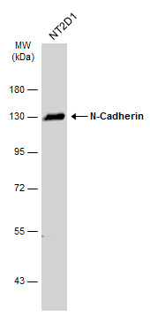
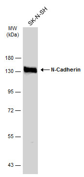
Whole cell extract (30 μg) was separated by 7.5% SDS-PAGE, and the membrane was blotted with N-Cadherin antibody (GRP520) diluted at 1:1000. The HRP-conjugated anti-rabbit IgG antibody was used to detect the primary antibody.
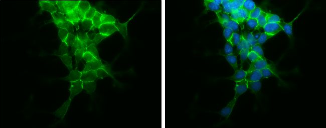
N-Cadherin antibody detects N-Cadherin protein at cell membrane by immunofluorescent analysis.Sample: SH-SY5Y cells were fixed in 4% paraformaldehyde at RT for 15 min.Green: N-Cadherin protein stained by N-Cadherin antibody (GRP520) diluted at 1:500.Blue:
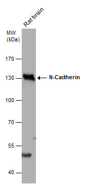
Rat tissue extract (50 μg) was separated by 7.5% SDS-PAGE, and the membrane was blotted with N-Cadherin antibody (GRP520) diluted at 1:1000. The HRP-conjugated anti-rabbit IgG antibody was used to detect the primary antibody.
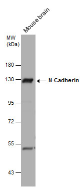
Mouse tissue extract (50 μg) was separated by 7.5% SDS-PAGE, and the membrane was blotted with N-Cadherin antibody (GRP520) diluted at 1:1000. The HRP-conjugated anti-rabbit IgG antibody was used to detect the primary antibody.
Whole cell extract (30 μg) was separated by 7.5% SDS-PAGE, and the membrane was blotted with N-Cadherin antibody (GRP520) diluted at 1:1000. The HRP-conjugated anti-rabbit IgG antibody was used to detect the primary antibody.
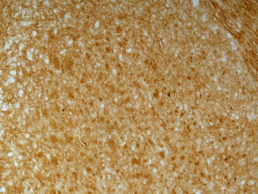
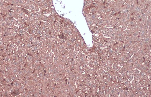
N-Cadherin antibody detects N-Cadherin protein at cell membrane and cytoplasm by immunohistochemical analysis.Sample: Paraffin-embedded mouse liver.N-Cadherin stained by N-Cadherin antibody (GRP520) diluted at 1:1000.Antigen Retrieval: Citrate buffer, pH
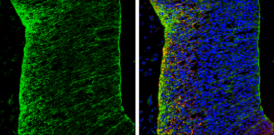
N-Cadherin antibody detects N-Cadherin protein expression by immunohistochemical analysis.Sample: Frozen sectioned E13.5 Rat brain. Green: N-Cadherin protein stained by N-Cadherin antibody (GRP520) diluted at 1:250.Red: beta Tubulin 3/ TUJ1, a mature neur
Reviews
Write Your Own Review
