Availability
- Request Lead Time
- In stock and ready for quick dispatch
- Usually dispatched within 5-10 working days
Product Overview
| Product Name | TET1 antibody [N1], N-term |
|---|---|
| Catalog Number | GRP64 |
| Species/Host | Rabbit |
| Reactivity | Human, Mouse |
| Conjugation | Unconjugated |
| Tested applications | ChIP, ICC, IF, IHC-P, WB |
| Immunogen | Recombinant protein encompassing a sequence within the N-terminus region of mouse TET1. The exact sequence is proprietary. |
| Alternative Names | (click to expand) |
Product Properties
| Form/Appearance | Liquid: 1XPBS, 20% Glycerol (pH7). 0.025% ProClin 300 was added as a preservative. |
|---|---|
| Concentration | 1 mg/ml |
| Storage | Store as concentrated solution. Centrifuge briefly prior to opening vial. For short-term storage (1-2 weeks), store at 4°C. For long-term storage, aliquot and store at -20°C or below. Avoid multiple freeze-thaw cycles. |
| Note | For research use only. |
| Isotype | IgG |
| Clonality | Polyclonal |
| Purity | Purified by antigen-affinity chromatography. |
| Uniprot ID | Q3URK3 |
| Entrez | 52463 |
Product Description
Dioxygenase that specifically binds methylcytosine (5mC), a minor base in mammalian DNA found in repetitive DNA elements that is crucial for retrotransposon silencing and mammalian development. Catalyzes the conversion of methylcytosine (5mC) to 5-hydroxymethylcytosine (hmC). The clear function of 5-hydroxymethylcytosine (hmC) is still unclear but it may influence chromatin structure and recruit specific factors or may constitute an intermediate component in cytosine demethylation. 5-hydroxymethylcytosine (hmC) is present in ES cells and is enriched in the brain, especially in Purkinje neurons. May play a role in the fetal development of heart, lung and brain.
Application Notes
| Dilution Range | WB: 1:500-1:20000,ICC: 1:100-1:1000 |
|---|
Validation Images
![TET1 antibody [N1], N-term detects TET1 protein at nucleus by immunofluorescent analysis.Sample: HepG2 cells were fixed in 4% paraformaldehyde at RT for 15 min.Green: TET1 protein stained by TET1 antibody [N1], N-term (GRP516) diluted at 1:500.Blue: Hoech](https://www.grp-ak.de/media/catalog/product/t/e/tet1-antibody-n1-n-term_grp516_if_1_2.jpg)
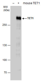
TET1 antibody detects TET1 protein by western blot analysis. Non-transfected (-) and mouse TET1-transfected (+, including DDDDK-tag) 293T whole cell extracts (30 μg) were separated by 5% SDS-PAGE, and the membrane was blotted with TET1 antibody (GRP516
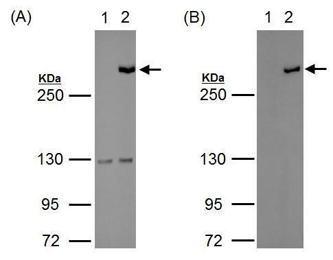
Western blot analysis of 293T cell are mock transfected (lane1) or with tagged-mTet1 (lane 2) for 24hrs, using either GRP516))) antibody. The HRP-conjugated anti-rabbit IgG antibody was used to detect the primary antibody.
TET1 antibody [N1], N-term detects TET1 protein at nucleus by immunofluorescent analysis.Sample: HepG2 cells were fixed in 4% paraformaldehyde at RT for 15 min.Green: TET1 protein stained by TET1 antibody [N1], N-term (GRP516) diluted at 1:500.Blue: Hoech
Reviews
Write Your Own Review
