Availability
- Request Lead Time
- In stock and ready for quick dispatch
- Usually dispatched within 5-10 working days
Product Overview
| Product Name | PD-L1 antibody |
|---|---|
| Catalog Number | GRP35 |
| Species/Host | Rabbit |
| Reactivity | Human |
| Conjugation | Unconjugated |
| Tested applications | ICC, IF, IHC-Fr, IHC-P, WB |
| Immunogen | Carrier-protein conjugated synthetic peptide encompassing a sequence within the C-terminus region of human PD-L1. The exact sequence is proprietary. |
| Alternative Names | (click to expand) |
Product Properties
| Form/Appearance | Liquid: 1XPBS, 1% BSA, 20% Glycerol (pH7). 0.025% ProClin 300 was added as a preservative. |
|---|---|
| Concentration | 0.24 mg/ml |
| Storage | Store as concentrated solution. Centrifuge briefly prior to opening vial. For short-term storage (1-2 weeks), store at 4°C. For long-term storage, aliquot and store at -20°C or below. Avoid multiple freeze-thaw cycles. |
| Note | For research use only. |
| Isotype | IgG |
| Clonality | Polyclonal |
| Purity | Purified by antigen-affinity chromatography. |
| Uniprot ID | Q9NZQ7 |
| Entrez | 29126 |
Product Description
Involved in the costimulatory signal, essential for T-cell proliferation and production of IL10 and IFNG, in an IL2-dependent and a PDCD1-independent manner. Interaction with PDCD1 inhibits T-cell proliferation and cytokine production.
Application Notes
| Dilution Range | WB: 1:500-1:3000,ICC: 1:100-1:1000,IHC-P: 1:100-1:1000 |
|---|
Validation Images
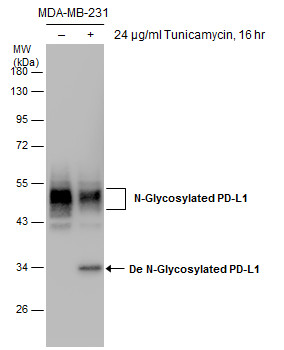
Untreated (–) and treated (+) MDA-MB-231 whole cell extracts (30 μg) were separated by 10% SDS-PAGE, and the membrane was blotted with PD-L1 antibody (GRP487) diluted at 1:1000.
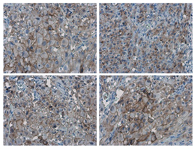
PD-L1 antibody detects PD-L1 proteinat cell membrane in human ovarian carcinoma by immunohistochemical analysis. Sample: Paraffin-embedded human ovarian carcinoma. PD-L1 antibody (GRP487) diluted at 1:1000.
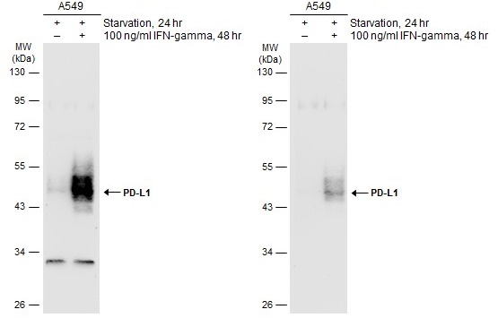
Untreated (–) and treated (+) A549 whole cell extracts (30 μg) were separated by 10% SDS-PAGE, and the membranes were blotted with PD-L1 antibody (GRP487) diluted at ) diluted at 1:500. The HRP-conjugated anti-rabbit IgG antibody was used to detect
PD-L1 antibody detects PD-L1 protein at cell membrane in human ovarian carcinoma by immunohistochemical analysis. Antibodies: PD-L1 antibody (GRP487) diluted at 1:1000, and competitor's antibody diluted at 1:50.
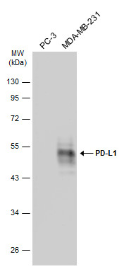
Various whole cell extracts (30 μg) were separated by 10% SDS-PAGE, and the membrane was blotted with PD-L1 antibody (GRP487) diluted at 1:2000. The HRP-conjugated anti-rabbit IgG antibody was used to detect the primary antibody, and the signal was de
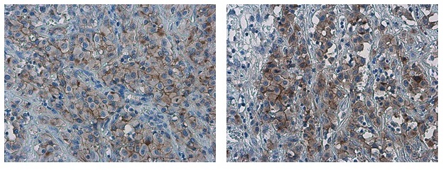
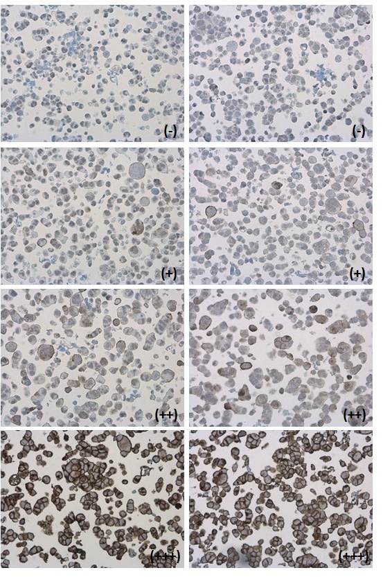
PD-L1 antibody detects PD-L1 protein at cell membrane in PD-L1 protein-expressing cell lines by immunohistochemical analysis. Antibodies: PD-L1 antibody (GRP487) diluted at 1:1000, and competitor's antibody diluted at 1:50. Samples: Negative (-), low po
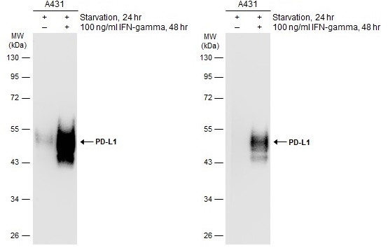
Untreated (–) and treated (+) A431 whole cell extracts (30 μg) were separated by 10% SDS-PAGE, and the membranes were blotted with PD-L1 antibody (GRP487) diluted at ) diluted at 1:500. The HRP-conjugated anti-rabbit IgG antibody was used to detect
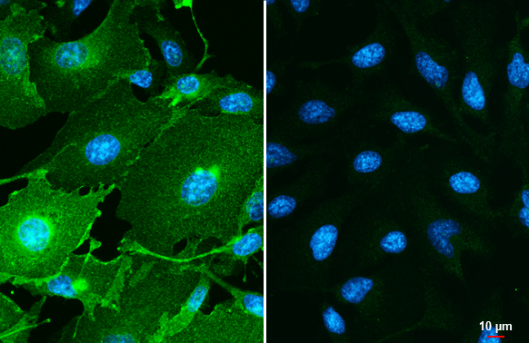
PD-L1 antibody detects PD-L1 protein by immunofluorescent analysis.Sample: MDA-MB-231 (left) and HeLa (right) cells were fixed in ice-cold MeOH for 5 min.Green: PD-L1 stained by PD-L1 antibody (GRP487) diluted at 1:500.Blue: Hoechst 33342 staining.Scale b
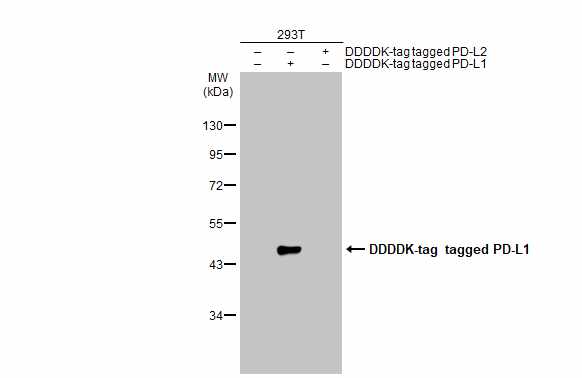
Non-transfected (–) and transfected (+) 293T whole cell extracts (30 μg) were separated by 10% SDS-PAGE, and the membrane was blotted with PD-L1 antibody (GRP487) diluted at 1:1000. The HRP-conjugated anti-rabbit IgG antibody was used to detect the
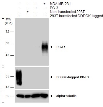
Various whole cell extracts were separated by 10% SDS-PAGE, and the membranes were blotted with PD-L1 antibody (GRP487) diluted at 1:600 and with DDDDK tag antibody (GRP487) diluted at 1:3000 to detect DDDDK-tagged PD-L2. The HRP-conjugated anti-rabbit Ig
Reviews
Write Your Own Review
