Availability
- Request Lead Time
- In stock and ready for quick dispatch
- Usually dispatched within 5-10 working days
Product Overview
| Product Name | NeuN antibody |
|---|---|
| Catalog Number | GRP164 |
| Species/Host | Rabbit |
| Reactivity | Mouse, Rat |
| Conjugation | Unconjugated |
| Tested applications | ICC, IF, IHC-Fr, IHC-P, WB |
| Immunogen | Carrier-protein conjugated synthetic peptide encompassing a sequence within the N-terminus region of human NeuN. The exact sequence is proprietary. |
| Alternative Names | (click to expand) |
Product Properties
| Form/Appearance | Liquid: 1XPBS, 20% Glycerol (pH7). 0.025% ProClin 300 was added as a preservative. |
|---|---|
| Concentration | 1.22 mg/ml |
| Storage | Store as concentrated solution. Centrifuge briefly prior to opening vial. For short-term storage (1-2 weeks), store at 4°C. For long-term storage, aliquot and store at -20°C or below. Avoid multiple freeze-thaw cycles. |
| Note | For research use only. |
| Isotype | IgG |
| Clonality | Polyclonal |
| Purity | Purified by antigen-affinity chromatography. |
| Uniprot ID | A6NFN3 |
| Entrez | 146713 |
Product Description
NeuN antibody
Application Notes
| Dilution Range | WB: 1:500-1:3000,ICC: 1:100-1:1000,IHC-P: 1:100-1:1000,IHC-Fr: 1:100-1:1000 |
|---|
Validation Images
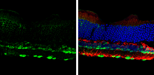
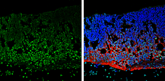
NeuN antibody detects NeuN protein by immunohistochemical analysis.Sample: Frozen sectioned adult mouse retina. Green: NeuN protein stained by NeuN antibody (GRP616) diluted at 1:250.Red: Protein kinase C alpha staining.Blue: Fluoroshield with DAPI.
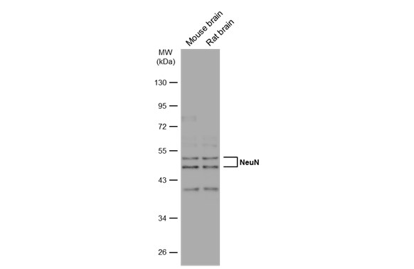
Various tissue extracts (50 μg) were separated by 10% SDS-PAGE, and the membrane was blotted with NeuN antibody (GRP616) diluted at 1:1000. The HRP-conjugated anti-rabbit IgG antibody was used to detect the primary antibody.
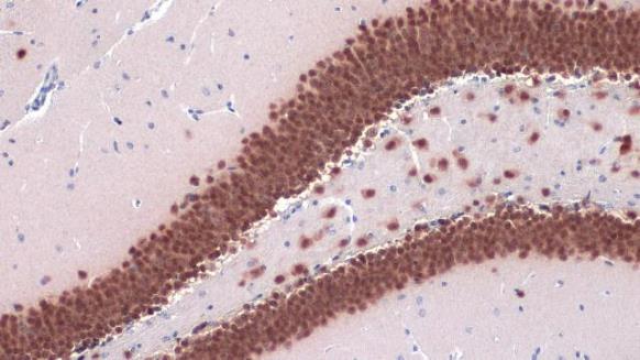
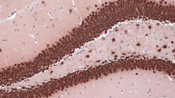
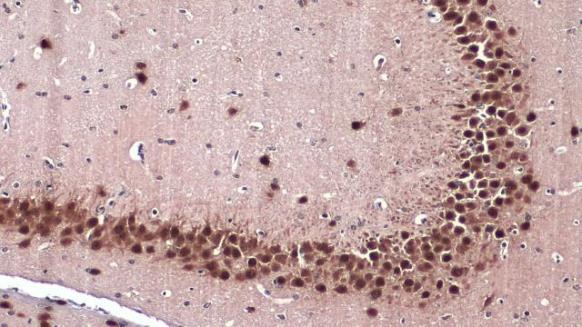
NeuN antibody detects NeuN protein at nucleus by immunohistochemical analysis.Sample: Paraffin-embedded mouse hippocampus.NeuN stained by NeuN antibody (GRP616) diluted at 1:2500.Antigen Retrieval: Citrate buffer, pH 6.0, 15 min
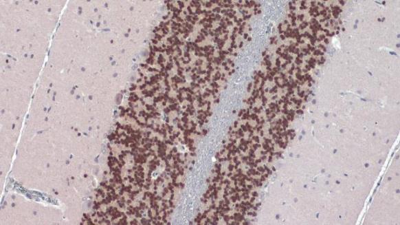
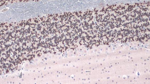
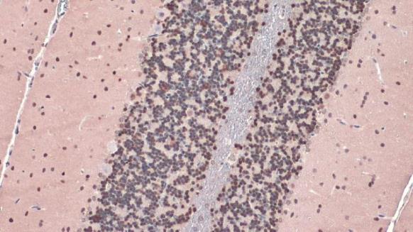
NeuN antibody detects NeuN protein at nucleus by immunohistochemical analysis.Sample: Paraffin-embedded rat cerebellum.NeuN stained by NeuN antibody (GRP616) diluted at 1:2500.Antigen Retrieval: Citrate buffer, pH 6.0, 15 min
NeuN antibody detects NeuN protein at nucleus by immunohistochemical analysis.Sample: Paraffin-embedded rat cerebellum.NeuN stained by NeuN antibody (GRP616) diluted at 1:1000.Antigen Retrieval: Citrate buffer, pH 6.0, 15 min
NeuN antibody detects NeuN protein at nucleus by immunohistochemical analysis.Sample: Paraffin-embedded mouse hippocampus.NeuN stained by NeuN antibody (GRP616) diluted at 1:1000.Antigen Retrieval: Citrate buffer, pH 6.0, 15 min
NeuN antibody detects NeuN protein at nucleus by immunohistochemical analysis.Sample: Paraffin-embedded mouse hippocampus.NeuN stained by NeuN antibody (GRP616) diluted at 1:500.Antigen Retrieval: Citrate buffer, pH 6.0, 15 min
NeuN antibody detects NeuN protein at nucleus by immunohistochemical analysis.Sample: Paraffin-embedded rat cerebellum.NeuN stained by NeuN antibody (GRP616) diluted at 1:500.Antigen Retrieval: Citrate buffer, pH 6.0, 15 min
NeuN antibody detects NeuN protein at nucleus by immunohistochemical analysis.Sample: Frozen sectioned E13.5 rat brain. Green: NeuN protein stained by NeuN antibody (GRP616) diluted at 1:250.Red: beta Tubulin 3/ TUJ1, a mature neuron marker, stained by be
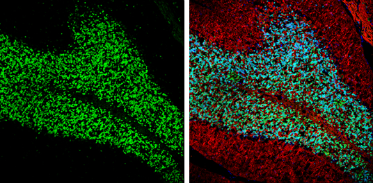
NeuN antibody detects NeuN protein expression by immunohistochemical analysis.Sample: Frozen-sectioned adult mouse cerebellum. Green: NeuN protein stained by NeuN antibody (GRP616) diluted at 1:250.Red: beta Tubulin 3/ TUJ1, stained by beta Tubulin 3/ TUJ
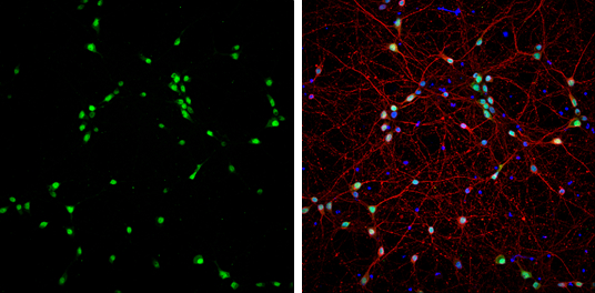
NeuN antibody detects NeuN protein at nucleus by immunofluorescent analysis.Sample: DIV9 rat E18 primary cortical neurons were fixed in 4% paraformaldehyde at RT for 15 min.Green: NeuN protein stained by NeuN antibody (GRP616) diluted at 1:1000.Red: beta
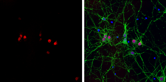
NeuN antibody detects NeuN protein at nucleus by immunofluorescent analysis.Sample: Rat E18 primary cortical neuron, DIV 8 cells were fixed in 4% paraformaldehyde at RT for 15 min.Red: NeuN protein stained by NeuN antibody (GRP616) diluted at 1:250.Green:
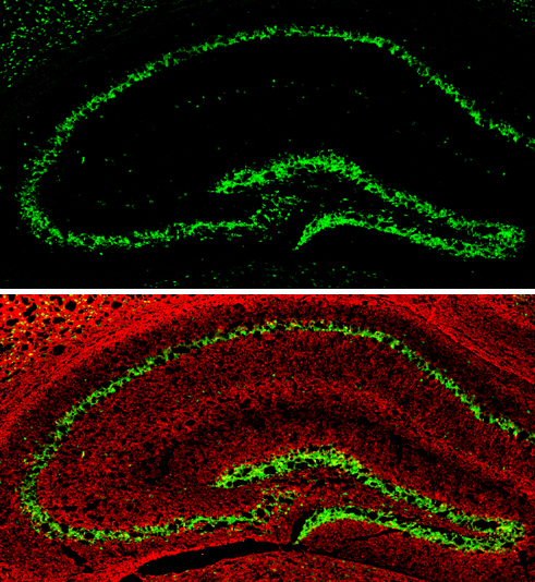
NeuN antibody detects NeuN protein expression by immunohistochemical analysis.Sample: Frozen-sectioned adult mouse hippocampus. Green: NeuN protein stained by NeuN antibody (GRP616) diluted at 1:250.Red: alpha Tubulin, stained by alpha Tubulin antibody [G
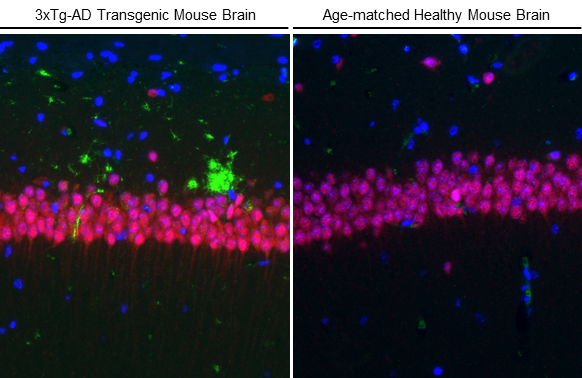
NeuN antibody detects NeuN protein at nucleus by immunohistochemical analysis.Sample: Paraffin-embedded 3xTg-AD transgenic mouse brain (left) and healthy mouse brain (right).Red: NeuN stained by NeuN antibody (GRP616) antibody – Conformation Specific an
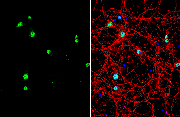
NeuN antibody detects NeuN protein by immunofluorescent analysis.Sample: DIV10 rat E18 primary cortical neuron cells were fixed in 4% paraformaldehyde at RT for 15 min.Green: NeuN stained by NeuN antibody (GRP616) diluted at 1:500.Red: Tau, stained by Tau
Reviews
Write Your Own Review
