Availability
- Request Lead Time
- In stock and ready for quick dispatch
- Usually dispatched within 5-10 working days
Product Overview
| Product Name | CBS antibody |
|---|---|
| Catalog Number | GRP139 |
| Species/Host | Rabbit |
| Reactivity | Human, Mouse, Rat |
| Conjugation | Unconjugated |
| Tested applications | ICC, IF, IHC-P, IP, WB |
| Immunogen | Recombinant protein encompassing a sequence within the center region of human CBS. The exact sequence is proprietary. |
| Alternative Names | (click to expand) |
Product Properties
| Form/Appearance | Liquid: 1XPBS, 20% Glycerol (pH7). 0.025% ProClin 300 was added as a preservative. |
|---|---|
| Concentration | 1 mg/ml |
| Storage | Store as concentrated solution. Centrifuge briefly prior to opening vial. For short-term storage (1-2 weeks), store at 4°C. For long-term storage, aliquot and store at -20°C or below. Avoid multiple freeze-thaw cycles. |
| Note | For research use only. |
| Isotype | IgG |
| Clonality | Polyclonal |
| Purity | Purified by antigen-affinity chromatography. |
| Uniprot ID | P35520|P0DN79 |
| Entrez | 875 |
Product Description
The protein encoded by this gene acts as a homotetramer to catalyze the conversion of homocysteine to cystathionine, the first step in the transsulfuration pathway. The encoded protein is allosterically activated by adenosyl-methionine and uses pyridoxal phosphate as a cofactor. Defects in this gene can cause cystathionine beta-synthase deficiency (CBSD), which can lead to homocystinuria. [provided by RefSeq]
Application Notes
| Dilution Range | WB: 1:500-1:3000,ICC: 1:100-1:1000,IHC-P: 1:100-1:1000,IP: 1:100-1:500 |
|---|
Validation Images
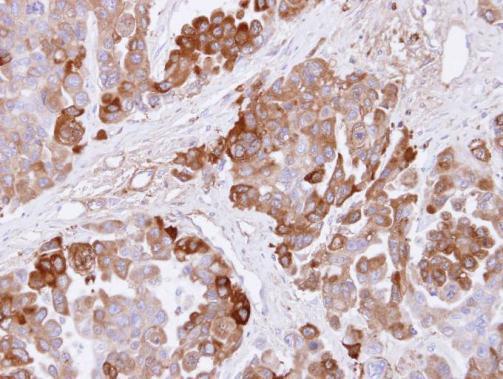
Immunohistochemical analysis of paraffin-embedded OVCAR3 xenograft , using CBS(GRP591) antibody at 1:500 dilution.
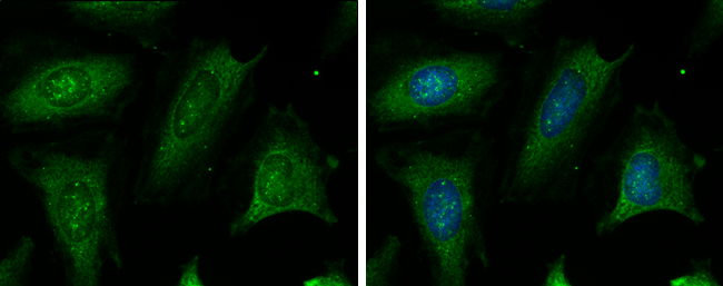
CBS antibody detects CBS protein at cytoplasm by immunofluorescent analysis.Sample: HeLa cells were fixed in ice-cold MeOH for 5 min.Green: CBS protein stained by CBS antibody (GRP591) diluted at 1:200.Blue: Hoechst 33342 staining.
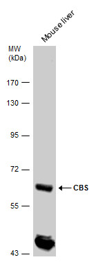
CBS antibody detects CBS protein by western blot analysis. Mouse tissue extracts (50 μg) was separated by 7.5% SDS-PAGE, and the membrane was blotted with CBS antibody (GRP591) diluted by 1:5000. The HRP-conjugated anti-rabbit IgG antibody was used to
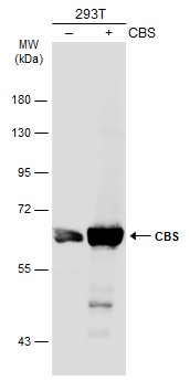
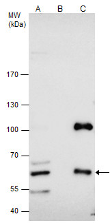
Non-transfected (–) and transfected (+) 293T whole cell extracts (30 μg) were separated by 7.5% SDS-PAGE, and the membrane was blotted with CBS antibody (GRP591) diluted at 1:1000. The HRP-conjugated anti-rabbit IgG antibody was used to detect the p
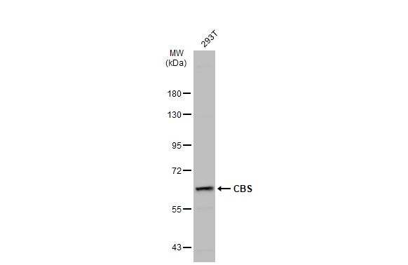
CBS antibody detects CBS protein by western blot analysis. Whole cell extracts (30 μg) was separated by 7.5% SDS-PAGE, and the membrane was blotted with CBS antibody (GRP591) at a dilution of 1:3000. The HRP-conjugated anti-rabbit IgG antibody was use
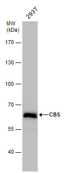
Whole cell extract (30 μg) was separated by 7.5% SDS-PAGE, and the membrane was blotted with CBS antibody (GRP591) diluted at 1:1000. The HRP-conjugated anti-rabbit IgG antibody was used to detect the primary antibody.
CBS antibody immunoprecipitates CBS protein in IP experiments.IP samples: HepG2 whole cell extractA. 50 ?g HepG2 whole cell extractB. Control with 4 ?g of preimmune Rabbit IgGC. Immunoprecipitation of CBS protein by 4 ?g CBS antibody (GRP591)7.5 % SDS-PAG
Reviews
Write Your Own Review
