Search results for: 'HRP antibody'
- 7 images
-
- 10 imagesAKT antibody [N3C2], Internal [GRP61]
ICC, IF, IHC-Fr, IHC-P, IP, WB
Human, Mouse, Rat, Fish
Rabbit
Polyclonal
100 μl -
- 4 images
-
- 1 imageMOUSE IgG (H&L) (GOAT) Antibody Peroxidase Conjugated (Min X Human Serum Proteins) [GRP4741]
ELISA, IHC-P, WB
Mouse
Goat
Polyclonal
100ug -
- 1 imageMOUSE IgG (H&L) (GOAT) Antibody Peroxidase Conjugated (Min X Human Serum Proteins) [GRP4740]
ELISA, IHC-P, WB
Mouse
Goat
Polyclonal
2mg -
-
-
-
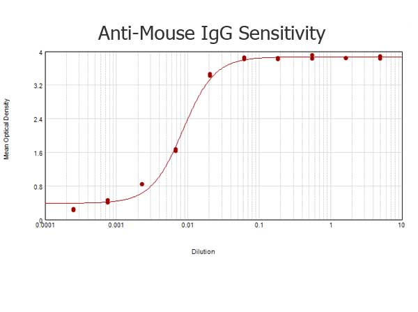
- Goat anti-Mouse IgG (H&L), HRP conjugated, min. cross-reactivity to human IgG or serum proteins [GRP13067]
Mouse
Goat
Polyclonal
1 mg - Donkey anti-Mouse IgG (H&L), F(ab)'2 Fragment, HRP conjugated [GRP13081]
FC, IL
Mouse
Donkey
Polyclonal
0.5 mg
![Non-transfected (–) and transfected (+) 293T whole cell extracts (30 μg) were separated by 10% SDS-PAGE, and the membrane was blotted with AKT antibody [N3C2], Internal (GRP513) diluted at 1:1000. The HRP-conjugated anti-rabbit IgG antibody was used](https://www.grp-ak.de/media/catalog/product/a/k/akt-antibody-n3c2-internal_grp513_wb_6_2.jpg)
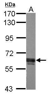
![Various whole cell extracts (30 μg) were separated by 7.5% SDS-PAGE, and the membrane was blotted with AKT antibody [N3C2], Internal (GRP513) diluted at 1:1000. The HRP-conjugated anti-rabbit IgG antibody was used to detect the primary antibody, and t](https://www.grp-ak.de/media/catalog/product/a/k/akt-antibody-n3c2-internal_grp513_wb_4_2.jpg)
![AKT antibody [N3C2], Internal detects AKT protein at cytoplasm by immunofluorescent analysis.Sample: HeLa cells were fixed in 4% paraformaldehyde at RT for 15 min.Green: AKT stained by AKT antibody [N3C2], Internal (GRP513) diluted at 1:500.Blue: Hoechst](https://www.grp-ak.de/media/catalog/product/a/k/akt-antibody-n3c2-internal_grp513_icc_1_2.jpg)
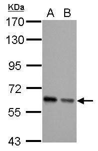
![Various whole cell extracts (30 μg) were separated by 10% SDS-PAGE, and the membrane was blotted with AKT antibody [N3C2], Internal (GRP513) diluted at 1:1000. The HRP-conjugated anti-rabbit IgG antibody was used to detect the primary antibody.](https://www.grp-ak.de/media/catalog/product/a/k/akt-antibody-n3c2-internal_grp513_wb_3_2.jpg)
![The WB analysis of AKT antibody [N3C2], Internal was published by Sun W and colleagues in the journal Cell Death Dis in 2014 .](https://www.grp-ak.de/media/catalog/product/a/k/akt-antibody-n3c2-internal_grp513_wb_2_2.jpg)
![The WB analysis of AKT antibody [N3C2], Internal was published by Vallejo-Flores G and colleagues in the journal Biomed Res Int in 2015.PMID: 26557697](https://www.grp-ak.de/media/catalog/product/a/k/akt-antibody-n3c2-internal_grp513_wb_1_2.jpg)
![Immunoprecipitation of Akt1/2/3 protein from 293T whole cell extracts using 5 ?g of Akt1/2/3 antibody [N3C2], Internal (GRP513).Western blot analysis was performed using Akt1/2/3 antibody [N3C2], Internal (GRP513).EasyBlot anti-Rabbit IgG was used as a s](https://www.grp-ak.de/media/catalog/product/a/k/akt-antibody-n3c2-internal_grp513_ip_1_2.jpg)