Search results for: 'proteinA'
- 12 imagesRad51 antibody [N1C2] [GRP8]
ICC, IF, IHC-P, IP, WB
Human, Mouse, Rat, Zebrafish
Rabbit
Polyclonal
100 μl -
- 9 imagesSQSTM1 / P62 antibody [N3C1], Internal [GRP15]
FACS, ICC, IF, IHC-P, IP, WB
Human, Mouse, Rat, Bovine, Zebrafish, Honeybee
Rabbit
Polyclonal
100 μl -
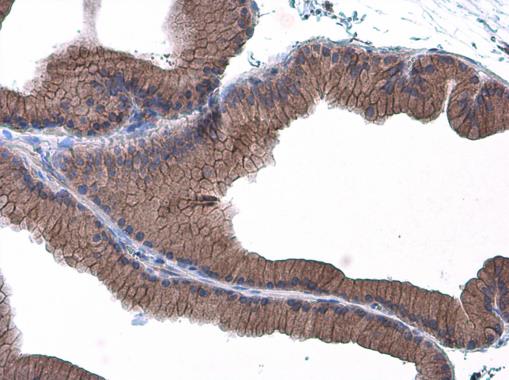
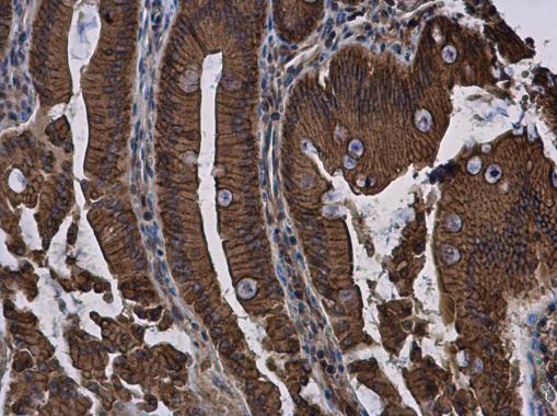
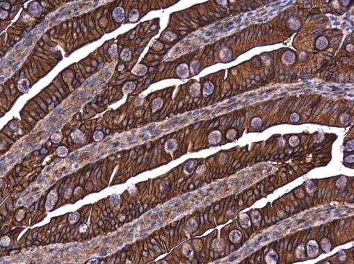
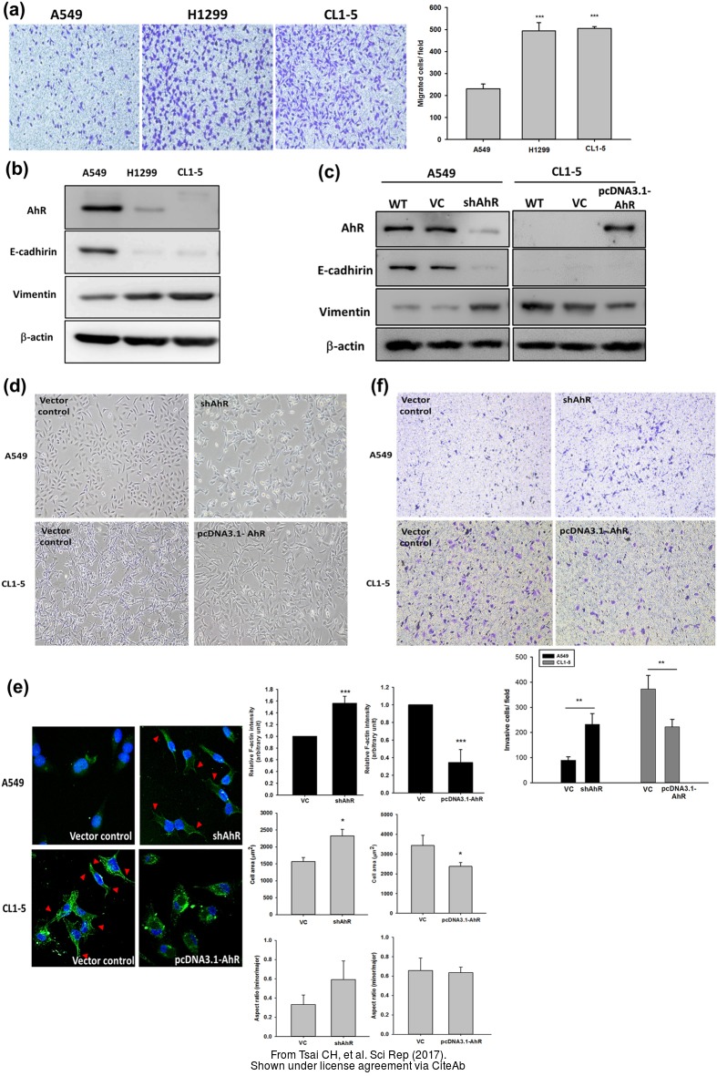
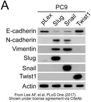
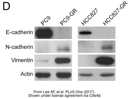
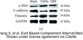
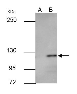
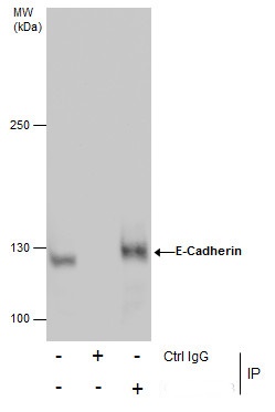
- 18 imagesE-Cadherin antibody [GRP7]
ICC, IF, IHC-P, IP, WB
Human, Mouse, Rat, Zebrafish
Rabbit
Polyclonal
100 μl -
![Various tissue extracts (30 μg) were separated by 10% SDS-PAGE, and the membrane was blotted with Rad51 antibody [N1C2] (GRP460) diluted at 1:500. The HRP-conjugated anti-rabbit IgG antibody was used to detect the primary antibody, and the signal was](https://www.grp-ak.de/media/catalog/product/r/a/rad51-antibody-n1c2_grp460_wb_7_2.jpg)
![Rad51 antibody [N1C2] detects Rad51 protein at nucleus by immunofluorescent analysis.Sample: HeLa cells were fixed in 4% paraformaldehyde at RT for 15 min.Green: Rad51 protein stained by Rad51 antibody [N1C2] (GRP460) diluted at 1:500.Red: phalloidin, a c](https://www.grp-ak.de/media/catalog/product/r/a/rad51-antibody-n1c2_grp460_if_1_2.jpg)
![Various whole cell extracts (30 μg) were separated by 10% SDS-PAGE, and the membrane was blotted with Rad51 antibody [14B4] (GRP460) diluted at 1:500. The HRP-conjugated anti-rabbit IgG antibody was used to detect the primary antibody.](https://www.grp-ak.de/media/catalog/product/r/a/rad51-antibody-n1c2_grp460_wb_6_2.jpg)
![Various whole cell extracts (30 μg) were separated by 10% SDS-PAGE, and the membrane was blotted with Rad51 antibody [N1C2] (GRP460) diluted at 1:1000.](https://www.grp-ak.de/media/catalog/product/r/a/rad51-antibody-n1c2_grp460_wb_5_2.jpg)
![Various whole cell extracts (30 μg) were separated by 10% SDS-PAGE, and the membrane was blotted with Rad51 antibody [14B4] (GRP460) diluted at 1:500. The HRP-conjugated anti-rabbit IgG antibody was used to detect the primary antibody.](https://www.grp-ak.de/media/catalog/product/r/a/rad51-antibody-n1c2_grp460_wb_4_2.jpg)
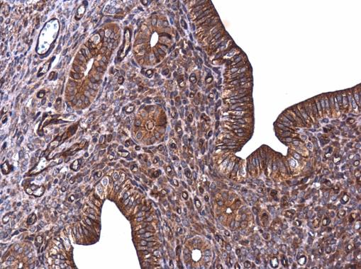
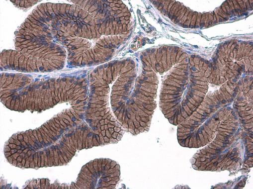
![Rad51 antibody [N1C2] detects Rad51 protein at cytoplasm and nucleus by immunohistochemical analysis.Sample: Paraffin-embedded human cervical carcinoma.Rad51 stained by Rad51 antibody [N1C2] (GRP460) diluted at 1:500.Antigen Retrieval: Citrate buffer, pH](https://www.grp-ak.de/media/catalog/product/r/a/rad51-antibody-n1c2_grp460_ihc-p_3_2.jpg)
![Rad51 antibody [N1C2] detects Rad51 protein at nucleus by immunohistochemical analysis.Sample: Paraffin-embedded mouse testis.Rad51 stained by Rad51 antibody [N1C2] (GRP460) diluted at 1:1000.Antigen Retrieval: Citrate buffer, pH 6.0, 15 min](https://www.grp-ak.de/media/catalog/product/r/a/rad51-antibody-n1c2_grp460_ihc-p_2_2.jpg)
![Rad51 antibody [N1C2] detects Rad51 protein at nucleus by immunohistochemical analysis.Sample: Paraffin-embedded mouse testis.Rad51 stained by Rad51 antibody [N1C2] (GRP460) diluted at 1:500.Antigen Retrieval: Citrate buffer, pH 6.0, 15 min](https://www.grp-ak.de/media/catalog/product/r/a/rad51-antibody-n1c2_grp460_ihc-p_1_2.jpg)
![Untreated (–) and treated (+) HeLa whole cell extracts (30 μg) were separated by 10% SDS-PAGE, and the membrane was blotted with Rad51 antibody [N1C2] (GRP460) diluted at 1:1000. The HRP-conjugated anti-rabbit IgG antibody was used to detect the pri](https://www.grp-ak.de/media/catalog/product/r/a/rad51-antibody-n1c2_grp460_wb_3_2.jpg)
![Various whole cell extracts (30 μg) were separated by 10% SDS-PAGE, and the membrane was blotted with Rad51 antibody [N1C2] (GRP460) diluted at 1:1000. The HRP-conjugated anti-rabbit IgG antibody was used to detect the primary antibody.](https://www.grp-ak.de/media/catalog/product/r/a/rad51-antibody-n1c2_grp460_wb_2_2.jpg)
![Various tissue extracts (30 μg) were separated by 10% SDS-PAGE, and the membrane was blotted with Rad51 antibody [N1C2] (GRP460) diluted at 1:1000. The HRP-conjugated anti-rabbit IgG antibody was used to detect the primary antibody, and the signal was](https://www.grp-ak.de/media/catalog/product/r/a/rad51-antibody-n1c2_grp460_wb_1_2.jpg)
![Rad51 antibody [N1C2] immunoprecipitates Rad51 protein in IP experiments.IP samples: Jurkat whole cell extractA. 40 ?g Jurkat whole cell extractB. Control with 4 ?g of preimmune Rabbit IgGC. Immunoprecipitation of Rad51 protein by 4 ?g Rad51 antibody [N1C](https://www.grp-ak.de/media/catalog/product/r/a/rad51-antibody-n1c2_grp460_ip_1_2.jpg)
![SQSTM1 antibody [N3C1], Internal (GRP467) detects SQSTM1 protein by flow cytometry analysis.Sample: HeLa cell fixed in 4% paraformaldehyde at 4ºC for 5 min.Brown: Unlabelled sample was also used as a control.Blue: SQSTM1 antibody [N3C1], Internal] dilut](https://www.grp-ak.de/media/catalog/product/s/q/sqstm1--p62-antibody-n3c1-internal_grp467_facs_1_2.jpg)
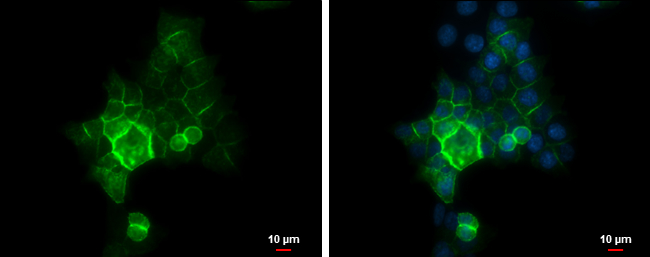
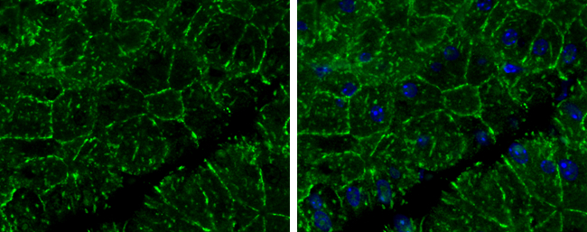
![SQSTM1 antibody [N3C1], Internal detects SQSTM1 protein at autophagosome by immunofluorescent analysis. Samples: HeLa cells mock (left) and treated with 50?M Chloroquine for 24 hr (right) were fixed in 4% paraformaldehyde at RT for 15 min.Green: SQSTM1 pr](https://www.grp-ak.de/media/catalog/product/s/q/sqstm1--p62-antibody-n3c1-internal_grp467_if_2_2.jpg)
![Untreated (–) and treated (+) HepG2 whole cell extracts (30 μg) were separated by 10% SDS-PAGE, and the membrane was blotted with SQSTM1 antibody [N3C1], Internal (GRP467) diluted at 1:1000. The HRP-conjugated anti-rabbit IgG antibody was used to de](https://www.grp-ak.de/media/catalog/product/s/q/sqstm1--p62-antibody-n3c1-internal_grp467_wb_4_2.jpg)
![SQSTM1 antibody [N3C1], Internal detects SQSTM1 protein by western blot analysis.A. 30 μg PC-12 whole cell lysate/extract B. 30 μg Rat2 whole cell lysate/extract10% SDS-PAGESQSTM1 antibody [N3C1], Internal (GRP467) dilution: 1:1000 The HRP-conjugate](https://www.grp-ak.de/media/catalog/product/s/q/sqstm1--p62-antibody-n3c1-internal_grp467_wb_3_2.jpg)
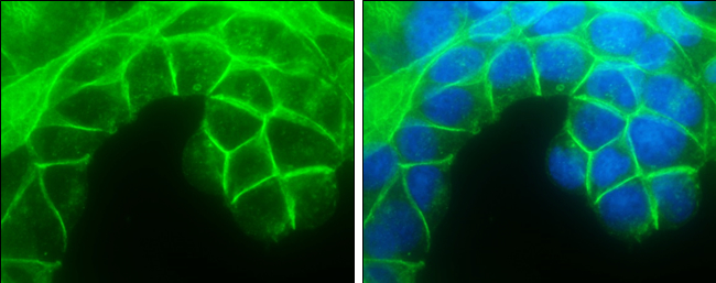
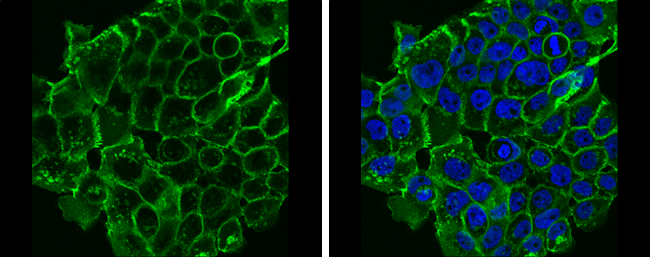
![SQSTM1 antibody [N3C1], Internal detects SQSTM1 protein at autophagosome by immunofluorescent analysis.Samples: HepG2 cells treated with 3?M thapsigargin 12 hrs (rigtht) and mock (left) were fixed in ice-cold MeOH for 10 min, permeabilize with cooled acet](https://www.grp-ak.de/media/catalog/product/s/q/sqstm1--p62-antibody-n3c1-internal_grp467_if_1_2.jpg)
![SQSTM1 antibody [N3C1], Internal detects SQSTM1 protein by western blot analysis.A. 30 μg NIH-3T3 whole cell lysate/extract B. 30 μg JC whole cell lysate/extract C. 30 μg BCL-1 whole cell lysate/extract 12% SDS-PAGESQSTM1 antibody [N3C1], Interna](https://www.grp-ak.de/media/catalog/product/s/q/sqstm1--p62-antibody-n3c1-internal_grp467_wb_2_2.jpg)
![Untreated (–) and treated (+) Huh-7 whole cell extracts (30 μg) were separated by 10% SDS-PAGE, and the membrane was blotted with SQSTM1 antibody [N3C1], Internal (GRP467) diluted at 1:1000. The HRP-conjugated anti-rabbit IgG antibody was used to de](https://www.grp-ak.de/media/catalog/product/s/q/sqstm1--p62-antibody-n3c1-internal_grp467_wb_1_2.jpg)
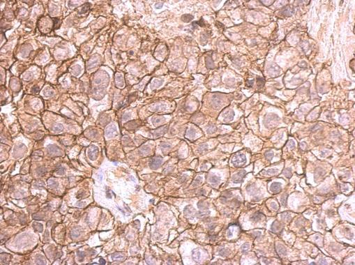
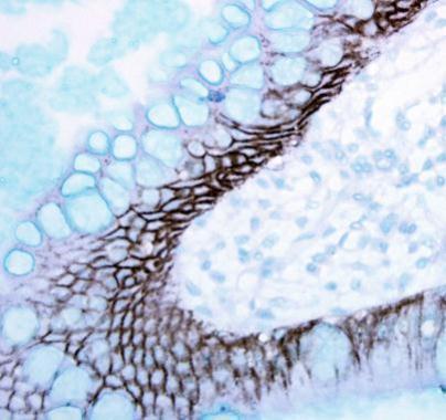
![SQSTM1 / P62 antibody [N3C1], Internal detects SQSTM1 / P62 protein at cytoplasm by immunohistochemical analysis.Sample: Paraffin-embedded human lung cancer.SQSTM1 / P62 stained by SQSTM1 / P62 antibody [N3C1], Internal (GRP467) diluted at 1:500.Antigen R](https://www.grp-ak.de/media/catalog/product/s/q/sqstm1--p62-antibody-n3c1-internal_grp467_ihc-p_1_2.jpg)
![Immunoprecipitation of SQSTM1 protein from HeLa whole cell extracts using 5 ?g of SQSTM1 antibody [N3C1], Internal (GRP467).Western blot analysis was performed using SQSTM1 antibody [N3C1], Internal (GRP467).EasyBlot anti-Rabbit IgG was used as a seconda](https://www.grp-ak.de/media/catalog/product/s/q/sqstm1--p62-antibody-n3c1-internal_grp467_ip_1_2.jpg)