Availability
- Request Lead Time
- In stock and ready for quick dispatch
- Usually dispatched within 5-10 working days
Product Overview
| Product Name | PGP9.5 antibody |
|---|---|
| Catalog Number | GRP126 |
| Species/Host | Rabbit |
| Reactivity | Human, Mouse, Rat |
| Conjugation | Unconjugated |
| Tested applications | ICC, IF, IHC-Fr, IHC-P, WB |
| Immunogen | Carrier-protein conjugated synthetic peptide encompassing a sequence within the C-terminus region of human PGP9.5. The exact sequence is proprietary. |
| Alternative Names | (click to expand) |
Product Properties
| Form/Appearance | Liquid: 1XPBS, 20% Glycerol (pH7). 0.025% ProClin 300 was added as a preservative. |
|---|---|
| Concentration | 1 mg/ml |
| Storage | Store as concentrated solution. Centrifuge briefly prior to opening vial. For short-term storage (1-2 weeks), store at 4°C. For long-term storage, aliquot and store at -20°C or below. Avoid multiple freeze-thaw cycles. |
| Note | For research use only. |
| Isotype | IgG |
| Clonality | Polyclonal |
| Purity | Purified by antigen-affinity chromatography. |
| Uniprot ID | P09936 |
| Entrez | 7345 |
Product Description
UCHL1 is a member of a gene family whose products hydrolyze small C-terminal adducts of ubiquitin to generate the ubiquitin monomer. Expression of UCHL1 is highly specific to neurons and to cells of the diffuse neuroendocrine system and their tumors. It is present in all neurons (Doran et al., 1983 [PubMed 6343558]).[supplied by OMIM]
Application Notes
| Dilution Range | WB: 1:1000-1:20000,ICC: 1:100-1:1000,IHC-P: 1:100-1:1000,IHC-Fr: 1:100-1:1000 |
|---|
Validation Images
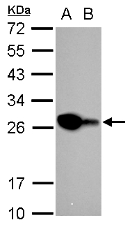
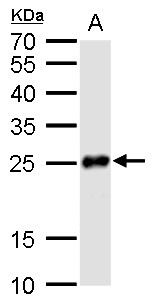
PGP9.5 antibody detects PGP9.5 protein by western blot analysis.A. 30 μg Neuro2A whole cell lysate/extractB. 30 μg GL261 whole cell lysate/extract12% SDS-PAGEPGP9.5 antibody (GRP578) dilution: 1:10000The HRP-conjugated anti-rabbit IgG antibody was
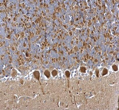
PGP9.5 antibody detects PGP9.5 protein at cytosol on rat hind brain by immunohistochemical analysis. Sample: Paraffin-embedded rat hind brain. PGP9.5 antibody (GRP578) dilution: 1:500.
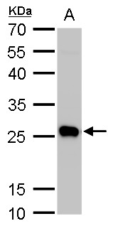
PGP9.5 antibody detects PGP9.5 protein by Western blot analysis.A. 50 ?g mouse brain lysate/extract12 % SDS-PAGEPGP9.5 antibody (GRP578) dilution: 1:5000
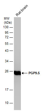
Rat tissue extract (50 μg) was separated by 12% SDS-PAGE, and the membrane was blotted with PGP9.5 antibody (GRP578) diluted at 1:10000.
PGP9.5 antibody detects PGP9.5 protein by Western blot analysis.A. 50 ?g rat brain lysate/extract12 % SDS-PAGEPGP9.5 antibody (GRP578) dilution: 1:10000
PGP9.5 antibody detects PGP9.5 protein by immunohistochemical analysis. Samples: Paraffin-embedded mouse skin.Green: PGP9.5 protein stained by PGP9.5 antibody (GRP578) diluted at 1:250.Red: beta Tubulin 3/ Tuj1, a marker, stained by beta Tubulin 3/ Tuj1
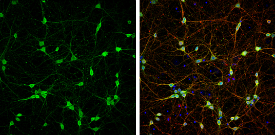
PGP9.5 antibody detects PGP9.5 protein at cytoplasm by immunofluorescent analysis.Sample: DIV9 rat E18 primary cortical neurons were fixed in 4% paraformaldehyde at RT for 15 min.Green: PGP9.5 protein stained by PGP9.5 antibody (GRP578) diluted at 1:500.R
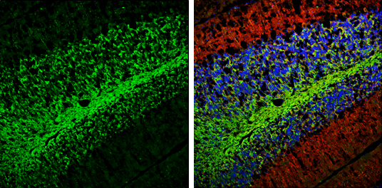
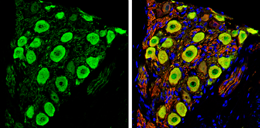
PGP9.5 antibody detects PGP9.5 protein expression by immunohistochemical analysis.Sample: Frozen-sectioned adult mouse cerebellum. Green: PGP9.5 protein stained by PGP9.5 antibody (GRP578) diluted at 1:250.Red: beta Tubulin 3/ TUJ1, stained by beta Tubuli
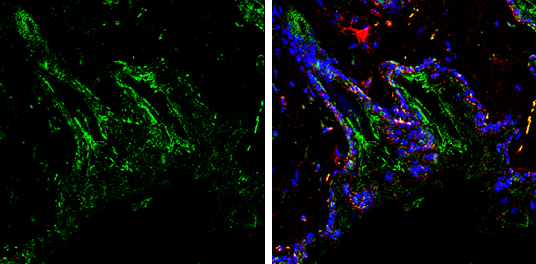
PGP9.5 antibody detects PGP9.5 protein by immunohistochemical analysis. Samples: Paraffin-embedded rat colon.Green: PGP9.5 protein stained by PGP9.5 antibody (GRP578) diluted at 1:250.Red: beta Tubulin 3/ Tuj1, a marker, stained by beta Tubulin 3/ Tuj1 a
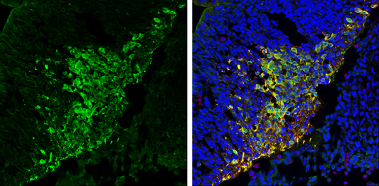
PGP9.5 antibody detects PGP9.5 protein expression by immunohistochemical analysis.Sample: Frozen sectioned E13.5 Rat brain. Green: PGP9.5 protein stained by PGP9.5 antibody (GRP578) diluted at 1:250.Red: beta Tubulin 3/ TUJ1, a mature neuron marker, stain
Reviews
Write Your Own Review
