Availability
- Request Lead Time
- In stock and ready for quick dispatch
- Usually dispatched within 5-10 working days
Product Overview
| Product Name | Rad51 antibody [14B4] |
|---|---|
| Catalog Number | GRP90 |
| Species/Host | Mouse |
| Reactivity | Human, Mouse, Rat, Chicken |
| Conjugation | Unconjugated |
| Tested applications | ICC, IF, IHC-P, IP, WB |
| Immunogen | Full length (amino acids 1-338) Rad51 expressed in E. coli. |
| Alternative Names | (click to expand) |
Product Properties
| Form/Appearance | Liquid: PBS |
|---|---|
| Concentration | 1.94 mg/ml |
| Storage | Store as concentrated solution. Centrifuge briefly prior to opening vial. For short-term storage (1-2 weeks), store at 4°C. For long-term storage, aliquot and store at -20°C or below. Avoid multiple freeze-thaw cycles. |
| Note | For research use only. |
| Isotype | IgG2b |
| Clonality | Monoclonal |
| Purity | Purified by antigen-affinity chromatography. |
| Clone ID | 14B4 |
| Uniprot ID | Q06609 |
| Entrez | 5888 |
Product Description
Rad51, a 37 kDa protein, is the human homologue of E. coli RecA protein and a member of the RAD52 epistasis group in S. cerevisiae. In recent studies it has been reported that BRCA1 interacts with Rad51 and disease-causing mutations have been found in the BRCA1 region necessary for BRCA1/Rad51 interaction, implying that this interaction is important for tumor suppression. BRCA2 and Rad51 have also been shown to interact by direct binding of the BRC repeats located within exon 11 of BRCA2. When these repeats are deleted, the interaction is lost and cells become hypersensitive to the DNA damage caused by methyl methanesulfonate (MMS).
Application Notes
| Dilution Range | WB: 1:500-1:3000,ICC: 1:100-1:1000,IHC-P: 1:100-1:1000 |
|---|
Validation Images
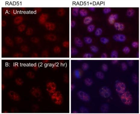
Immunofluorecent staning of RAD51 nuclear foci in U2OS cells using RAD51 14B4 antibody (GRP542). Cells were pre-extracted with CSK buffer before fixation with 4% PFA. RAD51 14B4 was used at 1:1000 dultion. DAPI was used to counterstain the nucleus. Scale
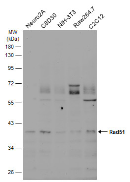
Various whole cell extracts (30 μg) were separated by 10% SDS-PAGE, and the membrane was blotted with Rad51 antibody [14B4] (GRP542) diluted at 1:500. The HRP-conjugated anti-mouset IgG antibody was used to detect the primary antibody, and the signal
Various whole cell extracts (30 μg) were separated by 10% SDS-PAGE, and the membrane was blotted with Rad51 antibody [14B4] (GRP542) diluted at 1:500. The HRP-conjugated anti-mouset IgG antibody was used to detect the primary antibody, and the signal
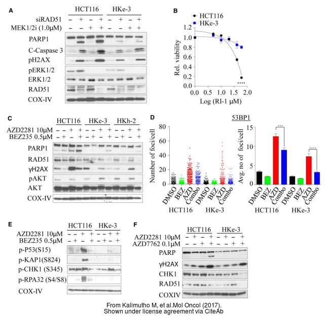
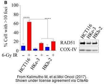
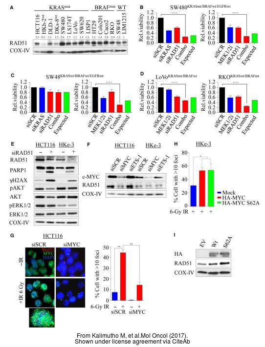
The WB analysis of Rad51 antibody [14B4] was published by Kalimutho M and colleagues in the journal Mol Oncol in 2017 .
The WB analysis of Rad51 antibody [14B4] was published by Kalimutho M and colleagues in the journal Mol Oncol in 2017 .
The WB analysis of Rad51 antibody [14B4] was published by Kalimutho M and colleagues in the journal Mol Oncol in 2017 .
The WB analysis of Rad51 antibody [14B4] was published by Kalimutho M and colleagues in the journal Mol Oncol in 2017 .
Various whole cell extracts (30 μg) were separated by 10% SDS-PAGE, and the membrane was blotted with Rad51 antibody [14B4] (GRP542) diluted at 1:500. The HRP-conjugated anti-mouse IgG antibody was used to detect the primary antibody, and the signal w
The WB analysis of Rad51 antibody [14B4] was published by Zhu J and colleagues in the journal EMBO Mol Med in 2013.PMID: 23341130
The WB analysis of Rad51 antibody [14B4] was published by Zhu J and colleagues in the journal EMBO Mol Med in 2013.PMID: 23341130
The ICC/IF analysis of Rad51 antibody [14B4] was published by White MK and colleagues in the journal PLoS One in 2014.PMID: 25310191
The WB analysis of Rad51 antibody [14B4] was published by Zhu J and colleagues in the journal EMBO Mol Med in 2013.PMID: 23341130
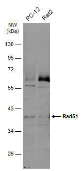
Whole cell extract (30 μg) was separated by 10% SDS-PAGE, and the membrane was blotted with Rad51 antibody [14B4] (GRP542) diluted at 1:500. The HRP-conjugated anti-mouse IgG antibody was used to detect the primary antibody.
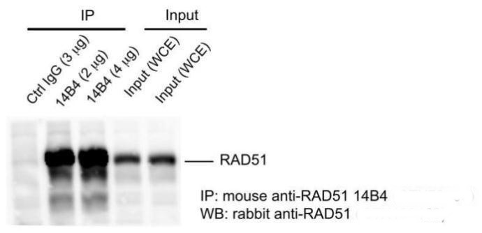
Mouse anti-RAD51 14B4 antibody (GRP542) was used in IP assay (immunoprecipitation) using HeLa cell extract prepared with lysis 180 buffer (40 mM Tris-HCl pH8.0, 180 mM NaCl, 1 mMEDTA, 0.5% NP-40).Rabbit anti-RAD51 antibody (GRP542) was used for subseque
Reviews
Write Your Own Review

![The WB analysis of Rad51 antibody [14B4] was published by Kalimutho M and colleagues in the journal Mol Oncol in 2017 .](https://www.grp-ak.de/media/catalog/product/r/a/rad51-antibody-14b4_grp542_wb_6_2.jpg)
![Various whole cell extracts (30 μg) were separated by 10% SDS-PAGE, and the membrane was blotted with Rad51 antibody [14B4] (GRP542) diluted at 1:500. The HRP-conjugated anti-mouse IgG antibody was used to detect the primary antibody, and the signal w](https://www.grp-ak.de/media/catalog/product/r/a/rad51-antibody-14b4_grp542_wb_5_2.jpg)
![The WB analysis of Rad51 antibody [14B4] was published by Zhu J and colleagues in the journal EMBO Mol Med in 2013.PMID: 23341130](https://www.grp-ak.de/media/catalog/product/r/a/rad51-antibody-14b4_grp542_wb_4_2.jpg)
![The WB analysis of Rad51 antibody [14B4] was published by Zhu J and colleagues in the journal EMBO Mol Med in 2013.PMID: 23341130](https://www.grp-ak.de/media/catalog/product/r/a/rad51-antibody-14b4_grp542_wb_3_2.jpg)
![The ICC/IF analysis of Rad51 antibody [14B4] was published by White MK and colleagues in the journal PLoS One in 2014.PMID: 25310191](https://www.grp-ak.de/media/catalog/product/r/a/rad51-antibody-14b4_grp542_icc_1_2.jpg)
![The WB analysis of Rad51 antibody [14B4] was published by Zhu J and colleagues in the journal EMBO Mol Med in 2013.PMID: 23341130](https://www.grp-ak.de/media/catalog/product/r/a/rad51-antibody-14b4_grp542_wb_2_2.jpg)
![Whole cell extract (30 μg) was separated by 10% SDS-PAGE, and the membrane was blotted with Rad51 antibody [14B4] (GRP542) diluted at 1:500. The HRP-conjugated anti-mouse IgG antibody was used to detect the primary antibody.](https://www.grp-ak.de/media/catalog/product/r/a/rad51-antibody-14b4_grp542_wb_1_2.jpg)
