Availability
- Request Lead Time
- In stock and ready for quick dispatch
- Usually dispatched within 5-10 working days
Product Overview
| Product Name | MAFG antibody |
|---|---|
| Catalog Number | GRP143 |
| Species/Host | Rabbit |
| Reactivity | Human, Mouse, Rat |
| Conjugation | Unconjugated |
| Tested applications | ChIP, ICC, IF, IHC-P, IP, WB |
| Immunogen | Recombinant protein encompassing a sequence within the center region of human MAFG. The exact sequence is proprietary. |
| Alternative Names | (click to expand) |
Product Properties
| Form/Appearance | Liquid: 1XPBS, 20% Glycerol (pH7). 0.025% ProClin 300 was added as a preservative. |
|---|---|
| Concentration | 0.53 mg/ml |
| Storage | Store as concentrated solution. Centrifuge briefly prior to opening vial. For short-term storage (1-2 weeks), store at 4°C. For long-term storage, aliquot and store at -20°C or below. Avoid multiple freeze-thaw cycles. |
| Note | For research use only. |
| Isotype | IgG |
| Clonality | Polyclonal |
| Purity | Purified by antigen-affinity chromatography. |
| Uniprot ID | O15525 |
| Entrez | 4097 |
Product Description
Globin gene expression is regulated through nuclear factor erythroid-2 (NFE2) elements located in enhancer-like locus control regions positioned many kb upstream of alpha- and beta-gene clusters (summarized by Blank et al., 1997 [PubMed 9166829]). NFE2 DNA-binding activity consists of a heterodimer containing a ubiquitous small Maf protein (MafF, MIM 604877; MafG; or MafK, MIM 600197) and the tissue-restricted protein p45 NFE2 (MIM 601490). Both subunits are members of the activator protein-1-like superfamily of basic leucine zipper (bZIP) proteins (see MIM 165160).[supplied by OMIM]
Application Notes
| Dilution Range | WB: 1:500-1:3000,ICC: 1:100-1:1000,IHC-P: 1:100-1:1000,IP: 1:100-1:500 |
|---|
Validation Images
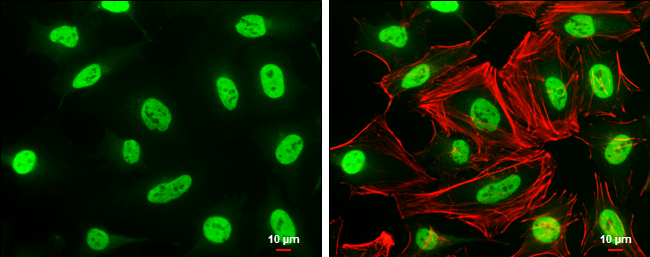
MAFG antibody detects MAFG protein at nucleus by immunofluorescent analysis.Sample: HeLa cells were fixed in 4% paraformaldehyde at RT for 15 min.Green: MAFG protein stained by MAFG antibody (GRP595) diluted at 1:500.Red: phalloidin, a cytoskeleton marker
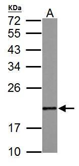
MAFG antibody detects MAFG protein by western blot analysis.A.50 μg rat muscle lysate/extract 12% SDS-PAGEMAFG antibody (GRP595) dilution: 1:1000 The HRP-conjugated anti-rabbit IgG antibody was used to detect the primary antibody.
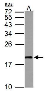
MAFG antibody detects MAFG protein by western blot analysis.A.50 μg mouse muscle lysate/extract 12% SDS-PAGEMAFG antibody (GRP595) dilution: 1:1000 The HRP-conjugated anti-rabbit IgG antibody was used to detect the primary antibody.
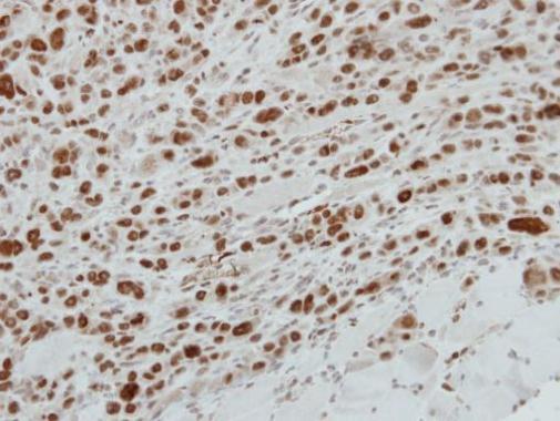
Immunohistochemical analysis of paraffin-embedded PC9 xenograft, using MAFG(GRP595) antibody at 1:500 dilution.
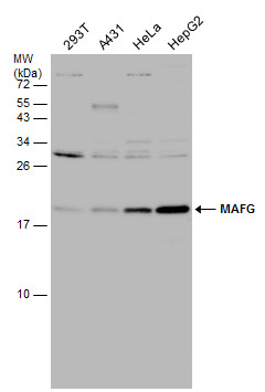
Various whole cell extracts (30 μg) were separated by 15% SDS-PAGE, and the membrane was blotted with MAFG antibody (GRP595) diluted at 1:1000.
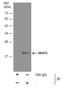
Immunoprecipitation of MAFG protein from HeLa whole cell extracts using 5 ?g of MAFG antibody (GRP595) .Western blot analysis was performed using MAFG antibody (GRP595) diluted at 1:500.EasyBlot anti-Mouse IgG was used as a secondary reagent.
Reviews
Write Your Own Review
