Availability
- Request Lead Time
- In stock and ready for quick dispatch
- Usually dispatched within 5-10 working days
Product Overview
| Product Name | Calbindin antibody |
|---|---|
| Catalog Number | GRP161 |
| Species/Host | Rabbit |
| Reactivity | Human, Mouse, Rat |
| Conjugation | Unconjugated |
| Tested applications | ICC, IF, IHC-Fr, IHC-P, WB |
| Immunogen | Recombinant protein encompassing a sequence within the center region of Human Calbindin. The exact sequence is proprietary. |
| Alternative Names | (click to expand) |
Product Properties
| Form/Appearance | Liquid: 1XPBS, 1% BSA, 20% Glycerol (pH7). 0.025% ProClin 300 was added as a preservative. |
|---|---|
| Concentration | 0.39 mg/ml |
| Storage | Store as concentrated solution. Centrifuge briefly prior to opening vial. For short-term storage (1-2 weeks), store at 4°C. For long-term storage, aliquot and store at -20°C or below. Avoid multiple freeze-thaw cycles. |
| Note | For research use only. |
| Isotype | IgG |
| Clonality | Polyclonal |
| Purity | Purified by antigen-affinity chromatography. |
| Uniprot ID | P05937 |
| Entrez | 793 |
Product Description
Calbindin is a calcium-binding protein belonging to the troponin C superfamily. It was originally described as a 27-kD protein induced by vitamin D in the duodenum of the chick. In the brain, its synthesis is independent of vitamin-D-derived hormones. Calbindin contains 4 active calcium-binding domains, and 2 modified domains that presumably have lost their calcium-binding capacity. The neurons in brains of patients with Huntington disease are calbindin-depleted. [provided by RefSeq]
Application Notes
| Dilution Range | WB: 1:500-1:3000,ICC: 1:100-1:1000,IHC-P: 1:100-1:1000,IHC-Fr: 1:100-1:1000 |
|---|
Validation Images
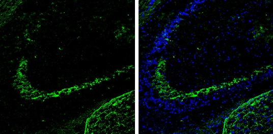
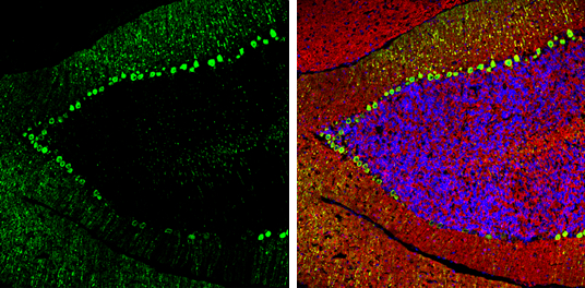
Calbindin antibody detects Calbindin protein expression by immunohistochemical analysis.Sample: Frozen-sectioned adult mouse hippocampus. Green: Calbindin protein stained by Calbindin antibody (GRP613) diluted at 1:250.Blue: Fluoroshield with DAPI.
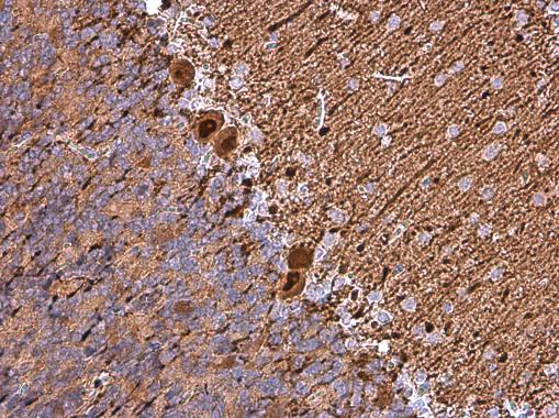
Calbindin antibody detects Calbindin protein at cytoplasm in rat brain by immunohistochemical analysis. Sample: Paraffin-embedded rat brain. Calbindin antibody (GRP613) diluted at 1:1000.
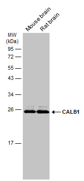
Various tissue extracts (50 μg) were separated by 12% SDS-PAGE, and the membrane was blotted with CALB1antibody (GRP613) diluted at 1:1000. The HRP-conjugated anti-rabbit IgG antibody was used to detect the primary antibody.
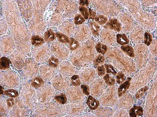
Calbindin antibody detects Calbindin protein at cytoplasm in mouse kidney by immunohistochemical analysis. Sample: Paraffin-embedded mouse kidney. Calbindin antibody (GRP613) diluted at 1:1000.
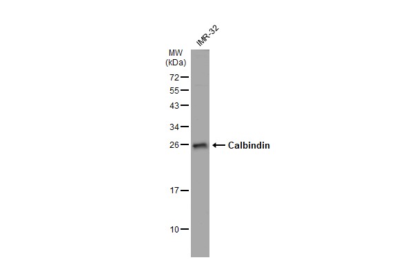
IMR-32 whole cell extracts (30 μg) was separated by 12% SDS-PAGE, and the membrane was blotted with Calbindin antibody (GRP613) diluted at 1:500. The HRP-conjugated anti-rabbit IgG antibody was used to detect the primary antibody, and the signal was d
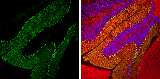
Calbindin antibody detects Calbindin protein expression by immunohistochemical analysis.Sample: Frozen-sectioned adult mouse cerebellum. Green: Calbindin protein stained by Calbindin antibody (GRP613) diluted at 1:250.Red: beta Tubulin 3/ TUJ1, stained by
Calbindin antibody detects Calbindin protein expression by immunohistochemical analysis.Sample: Frozen-sectioned adult mouse cerebellum. Green: Calbindin protein stained by Calbindin antibody (GRP613) diluted at 1:250.Red: beta Tubulin 3/ TUJ1, stained by
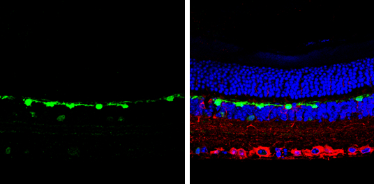
Calbindin antibody detects Calbindin protein by immunohistochemical analysis. Samples: Paraffin-embedded mouse retina.Green: Calbindin protein stained by Calbindin antibody (GRP613) diluted at 1:500.Red: beta Tubulin 3/ Tuj1, a marker, stained by beta Tu
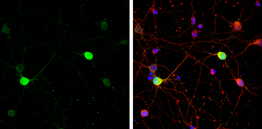
Calbindin antibody detects Calbindin protein by immunofluorescent analysis.Sample: DIV9 rat E18 primary cortical neuron cells were fixed in 4% paraformaldehyde at RT for 15 min.Green: Calbindin stained by Calbindin antibody (GRP613) diluted at 1:500.Red:
Reviews
Write Your Own Review
