Primary Antibodies
- 8 imagesCarbonic Anhydrase IX antibody [GT12] [GRP82]
FACS, ICC, IF, IHC-Fr, IHC-P, IP, WB
Human
Mouse
Monoclonal
100 μl -
- 12 imagesGlutamine synthetase antibody [GT1055] [GRP173]
ICC, IF, IHC-Fr, IHC-P, WB
Human, Mouse, Rat
Mouse
Monoclonal
100 μl -
- 10 imagesbeta Tubulin 3/ Tuj1 antibody [GT11710] [GRP174]
ICC, IF, IHC-Fr, IHC-P, IP, WB
Human, Mouse, Rat
Mouse
Monoclonal
100 μl -
- 7 imagesIba1 antibody [GT10312] [GRP175]
FACS, ICC, IF, IHC-Fr, IHC-P, WB
Human, Mouse, Rat
Mouse
Monoclonal
100 μl -
-
-
-
- Anti-Hu TCR gamma/delta Purified Low Endotoxin [GRP10384]
FC, FA, IHC-Fr, IHC-P
Human, Primate
Monoclonal
0.1 mg -
-
![Immunohistochemical analysis of paraffin-embedded cervical CA tissue sections using anti-CAIX antibody [GT12] (GRP534) at a dilution of 1:1000. The hypoxic regions of the tumor show positive CAIX staining.](https://www.grp-ak.de/media/catalog/product/c/a/carbonic-anhydrase-ix-antibody-gt12_grp534_ihc-p_5_2.jpg)
![Sample (30 μg HeLa whole cell lysate)A: 24 hr UntreatedB: 24 hr treatment with 100μM CoCl2C: 24 hr treatment with 200μM CoCl2D: 48 hr UntreatedE: 48 hr treatment with 100μM CoCl2F: 48 hr treatment with 200μM CoCl2Anti-CAIX antibody [GT12] (](https://www.grp-ak.de/media/catalog/product/c/a/carbonic-anhydrase-ix-antibody-gt12_grp534_wb_1_2.jpg)
![Immunohistochemical analysis of paraffin-embedded cervical CA tissue sections using anti-CAIX antibody [GT12] (GRP534) at a dilution of 1:1000. The hypoxic regions of the tumor show positive CAIX staining.](https://www.grp-ak.de/media/catalog/product/c/a/carbonic-anhydrase-ix-antibody-gt12_grp534_ihc-p_4_2.jpg)
![Confocal immunofluorescence analysis (Olympus FV10i) of methanol-fixed A431 cells treated with 200?M CoCl2 for 48hr using anti-CAIX antibody [GT12] (GRP534) at a dilution of 1:1000.](https://www.grp-ak.de/media/catalog/product/c/a/carbonic-anhydrase-ix-antibody-gt12_grp534_facs_2_2.jpg)
![Flow cytometry on HeLa cells (1x10^6) stained with anti-CAIX antibody [GT12] (GRP534) at a 1:1000 dilution. HeLa cells were untreated (green) or treated with 200?M CoCl2 (pink) for 48 hr.](https://www.grp-ak.de/media/catalog/product/c/a/carbonic-anhydrase-ix-antibody-gt12_grp534_facs_1_2.jpg)
![Immunohistochemical analysis of paraffin-embedded renal cell carcinoma (clear cell type) using anti-CAIX antibody [GT12] (GRP534) at a dilution of 1:1000.](https://www.grp-ak.de/media/catalog/product/c/a/carbonic-anhydrase-ix-antibody-gt12_grp534_ihc-p_3_2.jpg)
![Carbonic Anhydrase IX antibody [GT12] detects Carbonic Anhydrase IX protein at cell membrane by immunohistochemical analysis.Sample: Paraffin-embedded human cervical carcinoma.Carbonic Anhydrase IX stained by Carbonic Anhydrase IX antibody [GT12] (GRP534)](https://www.grp-ak.de/media/catalog/product/c/a/carbonic-anhydrase-ix-antibody-gt12_grp534_ihc-p_2_2.jpg)
![The IHC-P analysis of Carbonic Anhydrase IX antibody [GT12] was published by Huang WJ and colleagues in the journal PLoS One in 2015.PMID: 25738958](https://www.grp-ak.de/media/catalog/product/c/a/carbonic-anhydrase-ix-antibody-gt12_grp534_ihc-p_1_2.jpg)
![Glutamine synthetase antibody [GT1055] detects Glutamine synthetase protein on embryonic mouse brain by immunohistochemical analysis. Sample: Frozen section of embryonic mouse brain (mE18.5). Red: Glutamine synthetase antibody [GT1055] (GRP625) diluted at](https://www.grp-ak.de/media/catalog/product/g/l/glutamine-synthetase-antibody-gt1055_grp625_ihc_5_2.jpg)
![Glutamine synthetase antibody [GT1055] detects Glutamine synthetase protein at cytosol on human hepatoma by immunohistochemical analysis. Sample: Paraffin-embedded human hepatoma. Glutamine synthetase antibody [GT1055] (GRP625) dilution: 1:500.](https://www.grp-ak.de/media/catalog/product/g/l/glutamine-synthetase-antibody-gt1055_grp625_ihc_4_2.jpg)
![Non-transfected (–) and transfected (+) 293T whole cell extracts (30 μg) were separated by 10% SDS-PAGE, and the membrane was blotted with Glutamine synthetase antibody [GT1055] (GRP625) diluted at 1:1000. The HRP-conjugated anti-mouse IgG antibody](https://www.grp-ak.de/media/catalog/product/g/l/glutamine-synthetase-antibody-gt1055_grp625_wb_6_2.jpg)
![Glutamine synthetase antibody [GT1055] detects Glutamine synthetase protein by western blot analysis.A. 30 ?g 293T whole cell lysate/extract B. 30 ?g HeLa whole cell lysate/extract C. 30 ?g HepG2 whole cell lysate/extract10 % SDS-PAGEGlutamine synthetase](https://www.grp-ak.de/media/catalog/product/g/l/glutamine-synthetase-antibody-gt1055_grp625_wb_4_2.jpg)
![Glutamine synthetase antibody [GT1055] detects Glutamine synthetase protein at cytosol on mouse skin by immunohistochemical analysis. Sample: Paraffin-embedded mouse skin. Glutamine synthetase antibody [GT1055] (GRP625) dilution: 1:500.](https://www.grp-ak.de/media/catalog/product/g/l/glutamine-synthetase-antibody-gt1055_grp625_ihc_2_2.jpg)
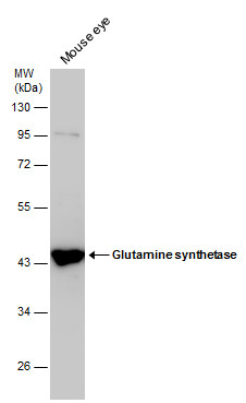
![Mouse tissue extract (50 μg) was separated by 10% SDS-PAGE, and the membrane was blotted with Glutamine synthetase antibody [GT1055] (GRP625) diluted at 1:5000.](https://www.grp-ak.de/media/catalog/product/g/l/glutamine-synthetase-antibody-gt1055_grp625_wb_3_2.jpg)
![Rat tissue extract (50 μg) was separated by 10% SDS-PAGE, and the membrane was blotted with Glutamine synthetase antibody [GT1055] (GRP625) diluted at 1:5000.](https://www.grp-ak.de/media/catalog/product/g/l/glutamine-synthetase-antibody-gt1055_grp625_wb_2_2.jpg)
![Glutamine synthetase antibody [GT1055] detects Glutamine synthetase protein at cytosol on rat hind brain by immunohistochemical analysis. Sample: Paraffin-embedded rat hind brain. Glutamine synthetase antibody [GT1055] (GRP625) dilution: 1:500.](https://www.grp-ak.de/media/catalog/product/g/l/glutamine-synthetase-antibody-gt1055_grp625_ihc_1_2.jpg)
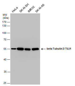
![Glutamine synthetase antibody [GT1055] detects Glutamine synthetase protein by western blot analysis.A. 30 ?g U87-MG whole cell lysate/extract B. 30 ?g SK-N-SH whole cell lysate/extract C. 30 ?g IMR32 whole cell lysate/extract D. 30 ?g SK-N-AS whole cell](https://www.grp-ak.de/media/catalog/product/g/l/glutamine-synthetase-antibody-gt1055_grp625_wb_1_2.jpg)
![Glutamine synthetase antibody [GT1055] detects Glutamine synthetase protein by immunohistochemical analysis.Sample: Frozen sectioned adult mouse retina.Green: Glutamine synthetase protein stained by Glutamine synthetase antibody [GT1055] (GRP625) diluted](https://www.grp-ak.de/media/catalog/product/g/l/glutamine-synthetase-antibody-gt1055_grp625_ihc_3_2.jpg)
![Glutamine synthetase antibody [GT1055] detects Glutamine synthetase protein at cytoplasm by immunofluorescent analysis.Sample: Cultured rat E18 primary cortical neuron, DIV 8. Cells were fixed in 4% paraformaldehyde at RT for 15 min.Green: Glutamine synth](https://www.grp-ak.de/media/catalog/product/g/l/glutamine-synthetase-antibody-gt1055_grp625_if_1_2.jpg)
![beta Tubulin 3/ TUJ1 antibody [GT11710] detects beta Tubulin 3/ TUJ1 protein by immunohistochemical analysis.Sample: Frozen sectioned E13.5 rat brain. Red: beta Tubulin 3/ TUJ1 protein stained by beta Tubulin 3/ TUJ1 antibody [GT11710] (GRP626) diluted at](https://www.grp-ak.de/media/catalog/product/b/e/beta-tubulin-3-tuj1-antibody-gt11710_grp626_ihc_4_2.jpg)
![beta III Tubulin antibody [GT11710] detects beta III Tubulin proteins on embryonic mouse brain by immunohistochemical analysis. Sample:Frozen section of embryonic mouse brain (mE18.5). Red: beta III Tubulin antibody [GT11710] (GRP626) diluted at 1:500. Bl](https://www.grp-ak.de/media/catalog/product/b/e/beta-tubulin-3-tuj1-antibody-gt11710_grp626_ihc_2_2.jpg)
![beta Tubulin 3/ TUJ1 antibody [GT11710] detects beta Tubulin 3/ TUJ1 protein by immunohistochemical analysis.Sample: Frozen sectioned adult mouse retina. Red: beta Tubulin 3/ TUJ1 protein stained by beta Tubulin 3/ TUJ1 antibody [GT11710] (GRP626) diluted](https://www.grp-ak.de/media/catalog/product/b/e/beta-tubulin-3-tuj1-antibody-gt11710_grp626_ihc_1_2.jpg)
![Various tissue extracts (10 μg) were separated by 10% SDS-PAGE, and the membrane was blotted with beta Tubulin 3/ Tuj1 antibody [GT11710] (GRP626) diluted at 1:20000. The HRP-conjugated anti-mouse IgG antibody was used to detect the primary antibody.](https://www.grp-ak.de/media/catalog/product/b/e/beta-tubulin-3-tuj1-antibody-gt11710_grp626_wb_3_2.jpg)
![beta Tubulin 3/ TUJ1 antibody [GT11710] detects beta Tubulin 3/ TUJ1 protein expression by immunofluorescent analysis.Sample: Cultured rat E18 primary hippocampal neuron. Cells were fixed in 4% paraformaldehyde at RT for 15 min.Green: beta Tubulin 3/ TUJ1](https://www.grp-ak.de/media/catalog/product/b/e/beta-tubulin-3-tuj1-antibody-gt11710_grp626_if_1_2.jpg)
![beta Tubulin 3/ TUJ1 antibody [GT11710] detects beta Tubulin 3/ TUJ1 protein at cytoplasm in rat brain by immunohistochemical analysis. Sample: Paraffin-embedded rat brain. beta Tubulin 3/ TUJ1 antibody [GT11710] (GRP626) diluted at 1:500.](https://www.grp-ak.de/media/catalog/product/b/e/beta-tubulin-3-tuj1-antibody-gt11710_grp626_ihc-p_1_2.jpg)
![Mouse tissue extract (30 μg) was separated by 10% SDS-PAGE, and the membrane was blotted with beta Tubulin 3/ Tuj1 antibody [GT11710] (GRP626) diluted at 1:5000. The HRP-conjugated anti-mouse IgG antibody was used to detect the primary antibody.](https://www.grp-ak.de/media/catalog/product/b/e/beta-tubulin-3-tuj1-antibody-gt11710_grp626_wb_1_2.jpg)
![beta Tubulin 3/ TUJ1 antibody [GT11710] detects beta Tubulin 3/ TUJ1 protein by immunohistochemical analysis.Sample: Frozen sectioned E13.5 rat brain.Green: SOX2 protein stained by SOX2 antibody [N1C3] (GRP626) diluted at 1:250.Red: beta Tubulin 3/ TUJ1 p](https://www.grp-ak.de/media/catalog/product/b/e/beta-tubulin-3-tuj1-antibody-gt11710_grp626_ihc_3_2.jpg)
![Immunoprecipitation of beta III Tubulin protein from SK-N-SH whole cell extracts using 5 ?g of beta III Tubulin antibody [GT11710] (GRP626).Western blot analysis was performed using beta III Tubulin antibody [GT11710] (GRP626).EasyBlot anti-Mouse IgG was](https://www.grp-ak.de/media/catalog/product/b/e/beta-tubulin-3-tuj1-antibody-gt11710_grp626_ip_1_2.jpg)
![Various whole cell extracts (30 μg) were separated by 15% SDS-PAGE, and the membrane was blotted with Iba1 antibody [GT10312] (GRP627) diluted at 1:500. The HRP-conjugated anti-mouse IgG antibody was used to detect the primary antibody.](https://www.grp-ak.de/media/catalog/product/i/b/iba1-antibody-gt10312_grp627_wb_2_2.jpg)
![Iba1 antibody [GT10312] detects Iba1 protein at cytoplasm by immunofluorescent analysis.Sample: THP-1 cells were fixed in 4% paraformaldehyde at RT for 15 min.Green: Iba1 protein stained by Iba1 antibody [GT10312] (GRP627) diluted at 1:200.Blue: Hoechst 3](https://www.grp-ak.de/media/catalog/product/i/b/iba1-antibody-gt10312_grp627_if_1_2.jpg)
![Whole cell extract (30 μg) was separated by 15% SDS-PAGE, and the membrane was blotted with Iba1 antibody [GT10312] (GRP627) diluted at 1:500. The HRP-conjugated anti-mouse IgG antibody was used to detect the primary antibody, and the signal was devel](https://www.grp-ak.de/media/catalog/product/i/b/iba1-antibody-gt10312_grp627_wb_1_2.jpg)
![Iba1 antibody [GT10312] detects Iba1 protein by immunohistochemical analysis.Sample: Frozen-sectioned mouse brain.Green: Iba1 stained by Iba1 antibody [GT10312] (GRP627) diluted at 1:200.Blue: Hoechst 33342 staining.](https://www.grp-ak.de/media/catalog/product/i/b/iba1-antibody-gt10312_grp627_ihc_1_2.jpg)
![Iba1 antibody [GT10312] detects Iba1 protein at cytoplasm by immunohistochemical analysis.Sample: Paraffin-embedded rat cerebellum.Iba1 stained by Iba1 antibody [GT10312] (GRP627) diluted at 1:1000.Antigen Retrieval: Citrate buffer, pH 6.0, 15 min](https://www.grp-ak.de/media/catalog/product/i/b/iba1-antibody-gt10312_grp627_ihc-p_2_2.jpg)
![Iba1 antibody [GT10312] detects Iba1 protein at cytoplasm by immunohistochemical analysis.Sample: Paraffin-embedded mouse cerebellum.Iba1 stained by Iba1 antibody [GT10312] (GRP627) diluted at 1:1000.Antigen Retrieval: Citrate buffer, pH 6.0, 15 min](https://www.grp-ak.de/media/catalog/product/i/b/iba1-antibody-gt10312_grp627_ihc-p_1_2.jpg)
![Iba1 antibody [GT10312] (GRP627) detects AIF1 protein by flow cytometry analysis. Sample: THP-1 cell. Black: Unlabelled sample was used as a control. Red: Iba1 antibody [GT10312] (GRP627) dilution: 1:50. Acquisition of 20,000 events were collected us](https://www.grp-ak.de/media/catalog/product/i/b/iba1-antibody-gt10312_grp627_facs_1_2.jpg)
